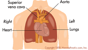
How Your Heart Works

The human heart.
Nucleus Medical Art, Inc./Getty Images

Ever­yone knows that the heart is a vital organ. We cannot live without our heart. However, when you get right down to it, the heart is just a pump. A complex and important one, yes, but still just a pump. As with all other pumps it can become clogged, break down and need repair. This is why it is critical that we know how the heart works. With a little knowledge about your heart and what is good or bad for it, you can significantly reduce your risk for heart disease.

Heart disease is the leading cause of death in the United States. Almost 2,000 Americans die of heart disease each day. That is one death every 44 seconds. The good news is that the death rate from heart disease has been steadily decreasing. Unfortunately, heart disease still causes sudden death and many people die before even reaching the hospital.

Advertisement

The heart holds a special place in our collective psyche as well. Of course the heart is synonymous with love. It has many other associations, too. Here are just a few examples:

  • have a heart - be merciful
  • change of heart - change your mind
  • to know something by heart - memorize something
  • broken heart - to lose love
  • heartfelt - deeply felt
  • have your heart in the right place - to be kind
  • cry your heart out - to grieve
  • heavy heart - sadness
  • have your heart set on - to want something badly

Certainly no other bodily organ elicits this kind of response. When was the last time you had a heavy pancreas?

In this article, we will look at this important organ so that you can understand exactly what makes your heart tick.

The Human Heart

[Please note - Medical illustrations assume that the patient is facing you so that the right and left correspond to the patient's right and left. That's why the left and right labels here seem backwards.]

The heart is a hollow, cone-shaped muscle located between the lungs and behind the sternum (breastbone). Two-thirds of the heart is located to the left of the midline of the body and 1/3 is to the right (see Figure 1).

The apex (pointed end) points down and to the left. It is 5 inches (12 cm) long, 3.5 inches (8-9 cm) wide and 2.5 inches (6 cm) from front to back, and is roughly the size of your fist. The average weight of a female human heart is 9 ounces and a male's heart is 10.5 ounces. The heart comprises less than 0.5 percent of the total body weight.

Advertisement

The heart has three layers. The smooth, inside lining of the heart is called the endocardium. The middle layer of heart muscle is called the myocardium. It is surrounded by a fluid filled sac call the pericardium.

Chambers and Valves

Figure 2
HowStuffWorks.com

The heart is divided into four chambers: (see Figure 2)

  1. right atrium (RA)
  2. right ventricle (RV)
  3. left atrium (LA)
  4. left ventricle (LV)

Each chamber has a sort of one-way valve at its exit that prevents blood from flowing backwards. When each chamber contracts, the valve at its exit opens. When it is finished contracting, the valve closes so that blood does not flow backwards.

Advertisement

  1. The tricuspid valve is at the exit of the right atrium.
  2. The pulmonary valve is at the exit of the right ventricle.
  3. The mitral valve is at the exit of the left atrium.
  4. The aortic valve is at the exit of the left ventricle.

When the heart muscle contracts or beats (called systole), it pumps blood out of the heart. The heart contracts in two stages. In the first stage, the right and left atria contract at the same time, pumping blood to the right and left ventricles. Then the ventricles contract together to propel blood out of the heart. Then the heart muscle relaxes (called diastole) before the next heartbeat. This allows blood to fill up the heart again.

The right and left sides of the heart have separate functions. The right side of the heart collects oxygen-poor blood from the body and pumps it to the lungs where it picks up oxygen and releases carbon dioxide. The left side of the heart then collects oxygen-rich blood from the lungs and pumps it to the body so that the cells throughout your body have the oxygen they need to function properly.

Blood Flow

All blood enters the right side of the heart through two veins: The superior vena cava (SVC) and the inferior vena cava (IVC) (see figure 3).

The SVC collects blood from the upper half of the body. The IVC collects blood from the lower half of the body. Blood leaves the SVC and the IVC and enters the right atrium (RA) (3).

Advertisement

When the RA contracts, the blood goes through the tricuspid valve (4) and into the right ventricle (RV) (5). When the RV contracts, blood is pumped through the pulmonary valve (6), into the pulmonary artery (PA) (7) and into the lungs where it picks up oxygen.

Why does it happen this way? Because blood returning from the body is relatively poor in oxygen. It needs to be full of oxygen before being returned to the body. So the right side of the heart pumps blood to the lungs first to pick up oxygen before going to the left side of the heart where it is returned to the body full of oxygen.

­ Blood now returns to the heart from the lungs by way of the pulmonary veins (8) and goes into the left atrium (LA) (9). When the LA contracts, blood travels through the mitral valve (10) and into the left ventricle (LV) (11). The LV is a very important chamber that pumps blood through the aortic valve (12) and into the aorta (13). The aorta is the main artery of the body. It receives all the blood that the heart has pumped out and distributes it to the rest of the body. The LV has a thicker muscle than any other heart chamber because it must pump blood to the rest of the body against much higher pressure in the general circulation (blood pressure).

Here is a recap of what we just discussed. Blood from the body flows:

  • to the superior and inferior vena cava,
  • then to the right atrium
  • through the tricuspid valve
  • to the right ventricle
  • through the pulmonic valve
  • to the pulmonary artery
  • to the lungs

The blood picks up oxygen in the lungs, and then flows from the lungs:

  • to the pulmonary veins
  • to the left atrium
  • through the mitral valve
  • to the left ventricle
  • through the aortic valve
  • to the aorta
  • to the body

The Body's Electrical System

Figure 4

Have you ever wondered what makes your heart beat? How does it do it automatically, every second of every minute of every hour of every day?

The answer lies in a special group of cells that have the ability to generate electrical activity on their own. These cells separate charged particles. Then they spontaneously leak certain charged particles into the cells. This produces electrical impuses in the pacemaker cells which spread over the heart, causing it to contract. These cells do this more than once per second to produce a normal heart beat of 72 beats per minute.

Advertisement

The natural pacemaker of the heart is called the sinoatrial node (SA node). It is located in the right atrium. The heart also contains specialized fibers that conduct the electrical impulse from the pacemaker (SA node) to the rest of the heart (see Figure 4).

The electrical impulse leaves the SA node (1) and travels to the right and left atria, causing them to contract together. This takes .04 seconds. There is now a natural delay to allow the atria to contract and the ventricles to fill up with blood. The electrical impulse has now traveled to the atrioventricular node (AV node) (2). The electrical impulse now goes to the Bundle of His (3), then it divides into the right and left bundle branches (4) where it rapidly spreads using Purkinje fibers (5) to the muscles of the right and left ventricle, causing them to contract at the same time.

Any of the electrical tissue in the heart has the ability to be a pacemaker. However, the SA node generates an electric impulse faster than the other tissue so it is normally in control. If the SA node should fail, the other parts of the electrical system can take over, although usually at a slower rate.

Although the pacemaker cells create the electrical impulse that causes the heart to beat, other nerves can change the rate at which the pacemaker cells fire and the how strongly the heart contracts. These nerves are part of the autonomic nervous system. The autonomic nervous system has two parts - The sympathetic nervous system and the parasympathetic nervous system. The sympathetic nerves increase the heart rate and increase the force of contraction. The parasympathetic nerves do the opposite.

All this activity produces electrical waves we can measure. The measurement is typically represented as a graph called an electrocardiogram (EKG). Here is an example of three heartbeats from an EKG (Figure 5):

Figure 5

Each part of the tracing has a lettered name:

  1. P wave - coincides with the spread of electrical activity over the atria and the beginning of its contraction.
  2. QRS complex - coincides with the spread of electrical activity over the ventricles and the beginning of its contraction.
  3. T wave - coincides with the recovery phase of the ventricles.

Electrical system abnormalities can range from minor premature beats (skipped beats) that do not require treatment, to slow or irregular beats that require an artificial pacemaker.

Blood Supply

Figure 6

Coronary arteries are the ones that we try to keep clear by eating a healthy diet. If coronary arteries are blocked a heart attack results.

The heart, just like any other organ, requires blood to supply it with oxygen and other nutrients so that it can do its work. The heart does not extract oxygen and other nutrients from the blood flowing inside it -- it gets its blood from coronary arteries that eventually carry blood within the heart muscle. Approximately 4 percent to 5 percent of the blood output of the heart goes to the coronary arteries (7 ½ ounces/minute or 225 ml/min).

Advertisement

There are two main coronary arteries (figure 6) - The left main coronary artery (1) and the right coronary artery (2) which arise from the aorta. The left main coronary artery divides into the left anterior descending branch (3) and the left circumflex arteries (4). Each artery supplies blood to different parts of the heart muscle and the electrical system.

The heart also has veins that collect oxygen-poor blood from the heart muscle. Most of the major veins of the heart (great cardiac vein, small cardiac vein, middle cardiac vein, posterior vein of the left ventricle, and oblique vein of the left atrium) drain into the coronary sinus which opens into the right atrium.

­ Coronary artery disease is caused by a blockage in one of the coronary arteries. When a coronary artery is partially blocked, that artery cannot supply enough blood to the heart muscle to meet its needs during exertion. When someone with coronary artery disease exerts himself or herself, it causes chest pain. This is due to lack of blood and oxygen to that part of the heart muscle and is called angina. If the obstruction worsens (more frequent angina episodes, with less exertion) a condition called unstable angina can occur. A heart attack happens when a coronary artery is completely blocked and no blood or oxygen is getting to the heart muscle served by that artery. This also causes chest pain and causes death to the heart muscle served by that artery.

For more information on the human heart and related topics, check out the links on the next page.