How does the spine form?

The human spine is shown here in rear and side views.
© iStockphoto.com/Raycat

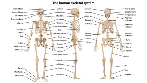
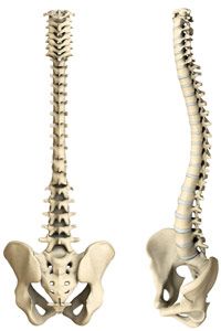
In a way, your spine is the keystone that holds your body together. It's at the center of your axial skeleton -- the flexible column that holds up the center of your body. At the top of your axial skeleton is your skull, and your coccyx, or tailbone, is at the other end. Together, all these bones shelter some of the most important organs in your body. Your brain sits in its protective skull casing, and the organs in your chest are protected by 12 pairs of ribs, all of which attach directly to your spine.

As if that wasn't enough, your spine is also what lets you move your arms and legs. Your arms attach to your spine and ribs via the collarbones and shoulder blades. Your legs attach to the spine through your hips. When you want to move your arms and legs, signals travel down your spinal cord, which is enclosed in your spine. Nerves carry the signals from the spinal cord to the muscles you want to move.

Advertisement

Arms and legs aren't the only parts of your body that move around, though -- your axial skeleton is also flexible. Discs provide lubrication between each of the 26 bones in your spine. As long as your discs are healthy, your vertebrae don't grind together when you bend and twist. Muscles attach to protrusions on the vertebrae called processes. When the muscles contract, they pull on these leverlike surfaces, and your vertebrae move.

Since your spine has a lot of responsibility, it's not surprising that it has to develop in exactly the right way in order for the whole system to work. So where does it come from, and what can go wrong as it grows?

Advertisement

Spinal Development

If you've read How Pregnancy Works, you already know about how a baby develops in a woman's body. A man's sperm joins with a woman's egg, creating a one-celled zygote. That one cell divides into two, which divide into four, over and over until there are about 100 cells. At this point, a few days after conception, the zygote becomes a blastocyst, and that's when the earliest beginnings of spinal development start to happen.

A blastocyst starts off as a collection of similar cells, but it doesn't stay that way for long. It develops three cell layers, called germ layers, in a process called gastrulation. The layers are the ectoderm, mesoderm and endoderm. Most of the body's innermost organs are formed from the endoderm, while most of the external features, like skin and hair, come from the ectoderm. The spine, part of the middle of the body, comes from the mesoderm. These layers are distinct within 12 days of the egg's fertilization.

Advertisement

The road from undifferentiated cells to a whole body is complex, so here's a rundown of what happens just in terms of the spine:

  1. The layers of the blastocyst move to where they're needed, arranging themselves to build a body.
  2. Cells from the mesoderm get together to form a structure called the notochord. This structure provides some support for the developing embryo.
  3. About 25 days after fertilization, the ectoderm above the notochord folds. The folds form a canal called the neural tube, which will become the central nervous system.
  4. Mesenchymal cells from the mesoderm form groups called somites on either side of the neural tube. These are like tiny building blocks that will become the vertebrae. Since an embryo develops a tail that disappears as it grows, there are more somites than vertebrae.
  5. When the embryo is 6 or 7 weeks old, ossification, or bone formation, begins. The somites harden, eventually becoming vertebrae. How Bones Work explains exactly what's happening during ossification. The notochord becomes part of the discs that lubricate the connections between the vertebrae.

In order for the spine to grow correctly, everything has to happen without a hitch, from migration of the blastocyst's layers to ossification. If the spinal column doesn't close correctly, the result can be one of a number of birth defects. Among the most common are neural tube defects, which include spina bifida and anencephaly, or a lack of brain development. Getting enough folate and folic acid during the earliest days of pregnancy reduces the risk of these defects.

You can learn more about the human body and related topics by following the links on the next page.

Advertisement

Lots More Information

Related HowStuffWorks Articles

More Great Links

  • Human Embryology
  • Spina Bifida Association

  • Tortora, Gerald J. and Sandra Reynolds Grabowsi. "Principles of Anatomy and Physiology." Ninth edition. John Wiley & Sons, Inc. New York. 2000.
  • Mayo Clinic. "Fetal Development: What happens in the first trimester?" 7/25/2007 (6/9/2009) http://www.mayoclinic.com/health/prenatal-care/PR00112
  • Medline Plus. "Neural Tube Defects." (6/9/2009) http://www.nlm.nih.gov/medlineplus/neuraltubedefects.html#cat1
  • Merck. "Brain and Spinal Cord Defects." Merck Manuals. (6/9/2009) http://www.merck.com/mmhe/sec23/ch265/ch265h.html
  • National Institute on Alcohol Abuse and Alcoholism. "Embryonic Development of the Nervous System." 2/2005. (6/9/2009) http://www.niaaa.nih.gov/Resources/GraphicsGallery/FetalAlcoholSyndrome/Embryonic.htm
  • Universities of Fribourg, Lausanne and Bern. "Human Embryology." Embryology.ch. (6/9/2009) http://www.embryology.ch/genericpages/moduleembryoen.html

Advertisement

Loading...