How Your Lungs Work

Transparent human chest showing heart and lungs
The heart and lungs work together to make sure your body has the oxygen-rich blood it needs. Callista Images/Getty Images

You breathe in and out anywhere from 15 to 25 times per minute without even thinking about it. When you exercise, your breathing rate goes up — again, without you thinking about it. You breathe so regularly that it is easy to take your lungs for granted. You can't even stop yourself from breathing if you try!

Lungs are the organs that help you breathe. They take a gas that your body needs to get rid of (carbon dioxide) and exchange it for a gas that your body can use (oxygen). They also work with your heart to make sure your body has the oxygen-rich blood it needs to function properly.

Advertisement

In this article, we will take a close look at how your lungs work and how they keep your body's cells supplied with oxygen and get rid of the carbon dioxide waste. We will explain some of the conditions and diseases that make breathing harder and cause the lungs to fail. We will also explain why you can't hold your breath for a long time and why you cough or hiccup.

How You Breathe

Your lungs are located within your chest cavity inside the rib cage (see illustration below). They are made of spongy, elastic tissue that stretches and constricts as you breathe. The airways that bring air into the lungs (the trachea and bronchi) are made of smooth muscle and cartilage, allowing the airways to constrict and expand. The lungs and airways bring in fresh, oxygen-enriched air and get rid of waste carbon dioxide made by your cells. They also help in regulating the concentration of hydrogen ion (pH) in your blood.

illustration of the lungs and diaphragm
When you inhale, the diaphragm and intercostal muscles (those are the muscles between your ribs) contract and expand the chest cavity.
HowStuffWorks

When you inhale, the diaphragm and intercostal muscles (those are the muscles between your ribs) contract and expand the chest cavity. This expansion lowers the pressure in the chest cavity below the outside air pressure. Air then flows in through the airways (from high pressure to low pressure) and inflates the lungs. When you exhale, the diaphragm and intercostal muscles relax and the chest cavity gets smaller. The decrease in volume of the cavity increases the pressure in the chest cavity above the outside air pressure. Air from the lungs (high pressure) then flows out of the airways to the outside air (low pressure). The cycle then repeats with each breath.

Advertisement

Where the Air Goes

As you breathe air in through your nose or mouth, it goes past the epiglottis and into the trachea. It continues down the trachea through your vocal cords in the larynx until it reaches the bronchi. From the bronchi, air passes into each lung. The air then follows narrower and narrower bronchioles until it reaches the alveoli.

illustration of red blood cell
At the beginning of the pulmonary capillary, the hemoglobin in the red blood cells has carbon dioxide bound to it and very little oxygen. The oxygen binds to hemoglobin and the carbon dioxide is released.
HowStuffWorks

Within each air sac, the oxygen concentration is high, so oxygen passes or diffuses across the alveolar membrane into the pulmonary capillary. At the beginning of the pulmonary capillary, the hemoglobin in the red blood cells has carbon dioxide bound to it and very little oxygen (see illustration above). The oxygen binds to hemoglobin and the carbon dioxide is released. Carbon dioxide is also released from sodium bicarbonate dissolved in the blood of the pulmonary capillary. The concentration of carbon dioxide is high in the pulmonary capillary, so carbon dioxide leaves the blood and passes across the alveolar membrane into the air sac. This exchange of gases occurs rapidly (fractions of a second). The carbon dioxide then leaves the alveolus when you exhale and the oxygen-enriched blood returns to the heart. Thus, the purpose of breathing is to keep the oxygen concentration high and the carbon dioxide concentration low in the alveoli so this gas exchange can occur!

Advertisement

Anatomy of the Lung

  • alveolus - tiny, thin-walled air sac at the end of the bronchiole branches where gas exchange occurs (plural - alveoli).
  • bronchioles - numerous small tubes that branch from each bronchus into the lungs. They get smaller and smaller.
  • bronchus - a branch of the trachea that goes from the trachea into the lung (plural - bronchi)
  • diaphragm - muscle at the base of the chest cavity that contracts and relaxes during breathing
  • epiglottis - a flap of tissue that closes over the trachea when you swallow so that food does not enter your airway
  • intercostal muscles - muscles along the rib cage that assist in breathing
  • larynx - voice box where the vocal cords are located.
  • nasal cavity - chamber in from the nose where air is moistened and warmed
  • pleural membranes - thin, membranes that cover the lungs, separate them from other organs and form a fluid-filled chest cavity.
  • pulmonary capillaries - small blood vessels that surround each alveolus
  • trachea -rigid tube that connects the mouth with the bronchi (windpipe)

Breathing and the Autonomic Nervous System

You don't have to think about breathing because your body's autonomic nervous system controls it, as it does many other functions in your body. If you try to hold your breath, your body will override your action and force you to let out that breath and start breathing again. The respiratory centers that control your rate of breathing are in the brainstem or medulla. The nerve cells that live within these centers automatically send signals to the diaphragm and intercostal muscles to contract and relax at regular intervals. However, the activity of the respiratory centers can be influenced by these factors:

  • Oxygen: Specialized nerve cells within the aorta and carotid arteries called peripheral chemoreceptors monitor the oxygen concentration of the blood and feed back on the respiratory centers. If the oxygen concentration in the blood decreases, they tell the respiratory centers to increase the rate and depth of breathing.
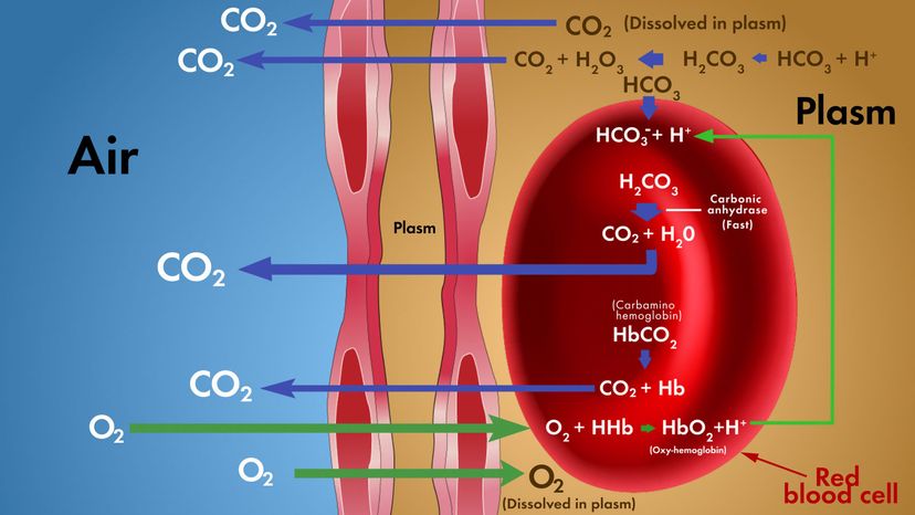
  • Carbon dioxide: Peripheral chemoreceptors also monitor the carbon dioxide concentration in the blood. In addition, a central chemoreceptor in the medulla monitors the carbon dioxide concentration in the cerebrospinal fluid (CSF) that surrounds the brain and spinal cord; carbon dioxide diffuses easily into the CSF from the blood. If the carbon dioxide concentration gets too high, then both types of chemoreceptors signal the respiratory centers to increase the rate and depth of breathing. The increased rate of breathing returns the carbon dioxide concentration to normal and the breathing rate then slows down.
  • Hydrogen ion (pH): The peripheral and central chemoreceptors are also sensitive to the pH of the blood and CSF. If the hydrogen ion concentration increases (that is, if the fluid becomes more acidic), then the chemoreceptors tell the respiratory centers to speed up. Hydrogen ion concentration is heavily influenced by the carbon dioxide concentration and bicarbonate concentration in the blood and CSF.
  • Stretch: Stretch receptors in the lungs and chest wall monitor the amount of stretch in these organs. If the lungs become over-inflated (stretch too much), they signal the respiratory centers to exhale and inhibit inspiration. This mechanism prevents damage to the lungs that would be caused by over-inflation.
  • Signals from higher brain centers: Nerve cells in the hypothalamus and cortex also influence the activity of the respiratory centers. During pain or strong emotions, the hypothalamus will tell the respiratory centers to speed up. Nerve centers in the cortex can voluntarily tell the respiratory center to speed up, slow down or even stop (holding your breath). Their influence, however, can be overridden by chemical factors (oxygen, carbon dioxide, pH).
  • Chemical irritants: Nerve cells in the airways sense the presence of unwanted substances in the airways such as pollen, dust, noxious fumes, water, or cigarette smoke. These cells then signal the respiratory centers to contract the respiratory muscles, causing you to sneeze or cough. Coughing and sneezing cause air to be rapidly and violently exhaled from the lungs and airways, removing the offending substance.

Of these factors, the strongest influence is the carbon dioxide concentration in your blood and CSF followed by the oxygen concentration.

Advertisement

Sometimes the respiratory centers go temporarily awry and send extra impulses to the diaphragm. These impulses cause unwanted contractions (hiccups). The same thing happens in unborn children; many pregnant women often feel their babies hiccup. This happens because the respiratory centers of the developing child's brain are working just like those of an adult even though they are not yet breathing air.

Lung Failure

There are many common conditions that can affect your lungs. Diseases or conditions of the lung fall mainly into two classes — those that make breathing harder and those that damage the lungs' ability to exchange carbon dioxide for oxygen.

Diseases or conditions that influence the mechanics of breathing:

Advertisement

  • Asthma: The bronchioles constrict, reducing the size of the airways. This cuts down on the flow of air and makes the respiratory muscles work harder.
  • Emphysema: The lungs become stiff with fibers and become less elastic, which increases the work of the respiratory muscles.
  • Bronchitis: The airways become inflamed and narrower, which restricts the flow of air and increases the work of the respiratory muscles
  • Pneumothorax: Air in the chest cavity equalizes the pressure in the chest cavity with the outside air and causes the lungs to collapse. This is usually caused by trauma or injury.
  • Apnea: Breathing slows or stops under a variety of conditions. There are many types of apnea, and they are usually caused by problems in the respiratory centers of the brain.

Diseases or conditions that minimize or prevent gas exchange:

  • Pulmonary edema: Fluid between the alveolus and pulmonary capillary builds up, which increases the distance over which gases must exchange and slows down the exchange.
  • Smoke inhalation: Smoke particles coat the alveoli and prevent the exchange of gases.
  • Carbon monoxide poisoning: Carbon monoxide binds to hemoglobin more tightly than either oxygen or carbon dioxide, which minimizes the delivery of oxygen to all the tissues of the body, including the brain, the heart and muscles. Carbon monoxide is a common product of poorly vented heaters (space heaters, furnaces, water heaters) and of automobile exhausts. This condition can be fatal if not caught soon after exposure.