How Therapeutic Hypothermia Works

Demonstration of therapeutic hypothermia
A research assistant chills out at the Hospital of the University of Pennsylvania during a demonstration of the therapeutic hypothermia procedure.
© Tim Shaffer/Reuters/Corbis

Whether you use that oatmeal jelly between your ears as a fine-tuned instrument of inquiry or as storage space for Monty Python quotes, you probably know that it works best when amply supplied with oxygen-rich blood. Your body, after all, is built from the cells upward for breathing and burning oxygen, and that goes double for neurons [source: Meek]. When this essential supply is cut off, a cascade of events affecting everything from your tissues to your cells is set in motion, the consequences of which can range from disability to death.

Over the past 70 years, medical research has revealed how refrigeration and resuscitation can go hand in hand, how otherwise life-threatening cold can be used to save lives and to delay this cycle of destruction.

Advertisement

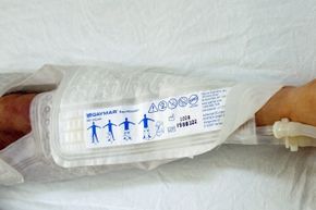
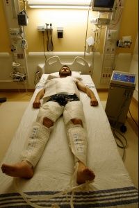
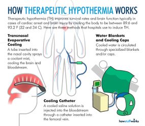
During therapeutic hypothermia (TH), also known as medically induced hypothermia or targeted temperature management, doctors lower oxygen demand in the brain by reducing a patient's body temperature to moderate hypothermic levels, thereby lowering the likelihood of cellular damage [sources: Deckard and Ebright; Gibson and Andrews]. Every 1.8 degrees Fahrenheit (1 degree Celsius) of body temperature reduction translates to an estimated 6-10 percent drop in cerebral metabolism and oxygen demand [sources: Adler at al.; Deckard and Ebright].

Typical procedures entail lowering core temperature past low normothermia (96.8-98.6 F, or 36-37 C) to a target range of 89.6-93.2 F (32-34 C) [sources: Deckard and Ebright; Gibson and Andrews]. That might not sound terribly frigid, but we're talking about core temperature, where a few degrees are all that separate a slight chill from heart and nervous system problems, organ damage and possible death [source: Davis].

In other words, don't try this at home.

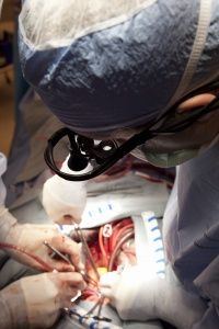
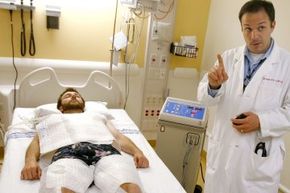
Through careful monitoring and temperature control, doctors can apply TH to cases that call for either intervention or prevention. Intervention involves curtailing the damage of a recent incident, one that deprived the brain of blood flow and oxygen. The two most common TH interventions involve cardiac arrest patients who don't wake up after circulation returns and newborns suffering from neonatal ischemic encephalopathy, an illness resulting from oxygen deprivation that can cause death, mental retardation, cerebral palsy and/or epilepsy, among other neurological effects [sources: Andrews; Deckard and Ebright; Lai and Yang; Leary; Merck Manual]. As a preventative measure, TH extends operating time and protects the brain during open heart surgery [source: Texas Heart Institute].

Over the past few decades, researchers have begun exploring using TH in treating specific types of stroke, heart attack, respiratory problems and injuries to the brain and spinal cord. Possible uses in treating acute kidney injury and managing cancer are in the early stages [sources: Encyclopedia Britannica; Leary].

Methods for inducing therapeutic hypothermia
© 2015 HowStuffWorks

Advertisement

Brief History of the Medical Ice Age

Open heart surgery
Many cardiac surgeons have embraced therapeutic hypothermia for decades.
© Dr. Barry Slaven/Visuals Unlimited/Corbis

The use of therapeutic hypothermia (TH) for improving outcomes in traumatic brain injury patients dates back to 1945, roughly the same year that Captain America became trapped in icy suspended animation [source: Gibson and Andrews].

Coincidence?

Advertisement

By the 1950s, open-heart surgeons were using TH as a preventative prelude to open-heart surgery. Although more recent techniques allow some cardiac surgeries to take place on a still-beating organ, historically such procedures have required stopping the heart, at least briefly. Researchers realized that hypothermia could help safeguard a patient's gray matter from the consequences of the resulting lack of blood flow (ischemia) and oxygen (anoxia) [sources: Nolan et al.; Texas Heart Institute]. Without TH, surgeons typically have less than five minutes to restore blood flow before brain damage begins to set in [source: Murphy].

Body cooling techniques are one of the two major technologies that make open heart surgeries possible. The other is the cardiopulmonary bypass machine (aka the heart-lung machine), which takes over oxygenation, carbon dioxide removal and blood pumping duties while the heart and lungs are inactive [sources: NHLBI; Texas Heart Institute].

As we'll discuss in greater detail below, TH has also seen use since the late 1950s in limiting damage to the central nervous systems of cardiac arrest patients who do not regain consciousness after the return of spontaneous circulation (ROSC) [sources: Deckard and Ebright; Gibson and Andrews]. Such benefits notwithstanding, TH's perceived ties to side effects like pneumonia, bleeding and cardiac arrhythmias left it medically sidelined until the 1990s, when experimental studies finally broke the ice by demonstrating that the technique could reduce neuronal damage and guard against cerebral ischemia [source: Gibson and Andrews].

Thereafter, research still moved at a somewhat glacial pace until the publication of two key cardiac studies in The New England Journal of Medicine in 2002. The so-called HACA (hypothermia after cardiac arrest) and Bernard studies both showed markedly improved outcomes in heart patients treated with hypothermia [sources: Deckard and Ebright; Winslow]. The studies' subjects suffered from either ventricular tachycardia, a rapid heartbeat spurred by offbeat electrical doings in the heart's lower chambers (ventricles), or ventricular fibrillation, a common heart arrhythmia in which ventricle muscles quiver haphazardly instead of performing a coordinated contraction [source: Deckard and Ebright].

In 2005, the American Heart Association (AHA) added therapeutic hypothermia to its cardiopulmonary resuscitation (CPR) guidelines [source: Deckard and Ebright; Winslow]. In 2010, they expanded these standards to include in-house cardiac arrests and patients incapable of following verbal commands after ROSC [source: Deckard and Ebright].

Uses and research continue to expand, but at its heart, TH provides a vital means of preserving the brain amid two of the harshest shocks it can suffer: the sudden loss of blood flow and its equally abrupt return.

Advertisement

Code Very, Very Blue

Although we often treat death as if it were a singular event, a moment in time separating human existence from What Comes After, in truth it is a complex terrain marked by vague milestones. Perhaps that's why death has so many definitions, from clinical death -- when heartbeat, breathing and circulation cease -- to the biological death that occurs sometime later, as brain cells begin to suffocate and we move beyond doctors' power to resuscitate.

The twilit country between those two borders is not a friendly one. In fact, it's under perpetual travel advisory, and with good reason. Take cardiac arrest. When it happens outside of a hospital, it kills 94 percent of the time, racking up an estimated 250,000 American deaths annually [source: Deckard and Ebright]. Those fortunate enough to survive face another grim prospect: a significant risk of developing neurological problems born not only of ischemia but also of reperfusion, the sudden return of blood flow following resuscitation [sources: Adler at al.; Deckard and Ebright]. Those injuries add to the biochemical, structural and functional problems that happen before and during the arrest. Thus, far from merely affecting the heart, cardiac arrest can entail a chain of cell destruction afflicting multiple organs, programmed cell death (apoptosis) in neurons and, possibly, bodily death [sources: Adler at al.; Deckard and Ebright].

Advertisement

Fortunately, many of these damaging processes are susceptible to low temperatures [sources: Adler at al.; Deckard and Ebright]. By cooling cardiac patients with signs of potential neurological issues after ROSC, the medical community has improved the odds of a positive neurological outcome from 20 percent to closer to 75 percent, with some patients reporting a full return to normalcy [sources: Deckard and Ebright; Winslow]. We've gone from a world in which 6-10 minutes with no pulse meant inevitable brain death to one in which that time can be stretched to 20 minutes [source: Winslow]. More radical experimental approaches hold the promise of extending the resuscitation window even longer (see sidebar).

Resuscitation. That's an important word. Because, chilled or not, patents with no pulse, breathing or circulation are dead in a way that will trigger most Do Not Resuscitate orders.

So what's happening here? How do a few degrees of lowered body temperature measure the gap between bad and much, much worse? To understand that, we need to see what goes on when your heart shuts off.

Advertisement

Chain of Death

Mitochondria in the human heart
© Visuals Unlimited/Corbis

Our organs experience significant injuries not only when oxygen-supplying blood flow is curtailed, but also when it suddenly returns. Mitigating the neurological effects of this ischemia-reperfusion injury (aka post-cardiac arrest syndrome) is one of therapeutic hypothermia's primary applications [source: Delfin].

When blood and oxygen flow to the brain cease, a cascade of biochemical events kicks off, one that can continue to destroy brain cells for hours or days after circulation is restored [source: Delfin]. One of the most pivotal aspects of this process concerns mitochondria, the vital, sausage-shaped organelles that power our cells.

Advertisement

Without oxygen, mitochondria's normal energy processes fail, and cells switch to anaerobic respiration. This causes lactic acid to build up, essential mechanisms to shut down and calcium to accumulate within the cell. The resulting glut of calcium prompts the discharge of the neurotransmitter glutamate, which excites brain cells, sparking a more urgent call for nonexistent oxygen and triggering a whole host of harmful chemicals, including highly reactive free radicals [sources: Deckard and Ebright; Delfin; Merck Manual].

Meanwhile, cell walls lose integrity and let in additional harmful substances, including more calcium, as well as sodium, which causes inflammation (edema). Overcome, the mitochondria die and break down, releasing toxins and other chemicals that can trigger apoptosis. If the cell then kicks the bucket, it releases glutamate and toxins into its surroundings, exciting nearby neurons. The immune system dispatches neutrophils and macrophages to sweep up dead cells, kicking off a chain reaction by producing free radicals that further damage cells, deepening the inflammatory response, aggravating cerebral swelling and causing further neurological injury. Meanwhile, the combination of ischemia, swelling and pressure cause the protective blood-brain barrier to grow more permeable [sources: Deckard and Ebright; Gibson and Andrews].

That's the bad news. The worse news is that when circulation resumes it further damages these already weakened tissues and cells. Leaky cell membranes and a destabilized blood-brain barrier mean further cerebral edema as newly delivered white blood cells gather in the brain and, provoked by damaged tissue, let loose a horde of inflammatory factors and free radicals. Meanwhile, blood coagulation and the formation of microclots can block blood flow, causing localized ischemia. Seizure activity can further influence the course of such complications [source: Delfin].

This is, of course, a vastly simplified overview, but it reveals the harmful mechanisms that therapeutic hypothermia helps to shut down.

Advertisement

You Should Put Some Ice on That

Demonstration of therapeutic hypothermia
A research assistant from the Hospital of the University of Pennsylvania demonstrates how a patient receiving therapeutic hypothermia might have his or her leg wrapped.
© Tim Shaffer/Reuters/Corbis

In the previous section, we described the biochemical rioting and looting that sweeps through your brain cells when oxygen is lacking, and the further injury that accumulates after blood flow returns. We also identified a rogue's gallery of troublemakers: starving mitochondria, wandering electrolytes, destructive enzymes, programmed cell death, runaway neuron excitability, unwanted inflammation and poorly timed clotting.

Therapeutic hypothermia blunts the force of all of these factors. First and foremost, it slows the cerebral metabolic rate, thereby reducing oxygen demand by brain cells and staving off mitochondrial meltdown [sources: Adler at al.; Delfin; Gibson and Andrews; Winslow]. By stabilizing glutamate release, it also put the brakes on damage-boosting cell excitation [sources: Adler et al.; Deckard and Ebright; Delfin; Merck Manual]. TH also shores up the blood-brain barrier by making small blood vessels called arterioles less leaky [sources: Deckard and Ebright; Gibson and Andrews].

Advertisement

Remember how runaway calcium levels generally wrecked brain cells and set off a self-feeding chain reaction? Hypothermia calms that down too, thereby lowering the rate of mitochondria damage, cell death and all that typically follows, including destructive free radical production and brain inflammation [sources: Adler et al.; Deckard and Ebright; Delfin; Gibson and Andrews; Merck Manual]. TH also reduces the inflammation response by tamping down on the release of cell-signaling proteins called cytokines, which can contribute to clotting, blood vessel breakdown and cell death [sources: Adler at al.; Delfin; Gibson and Andrews; Merck Manual]. Finally, studies have shown that therapeutic hypothermia reduces seizure activity, an important factor governing the chances of positive neurological recovery [source: Gibson and Andrews].

But don't take the polar plunge just yet. There are good reasons our bodies try to maintain a constant temperature. More to the point, therapeutic hypothermia comes with a number of side effects that might occur during the cooling, maintenance and rewarming stages of the process. These can include shivering, cardiovascular problems, mild coagulation problems, issues with blood glucose, electrolyte imbalances and, rarely, pneumonia [sources: Deckard and Ebright; Gibson and Andrews]. During warming, the patent may experience a rise in intracranial pressure and, occasionally (and somewhat ironically), hyperthermia (elevated body temperature) [source: Adler at al.].

Consequently, TH requires careful monitoring of a variety of vital signs during both the procedure and recovery. Let's take a closer look at the actual procedure of therapeutic hypothermia to see how this plays out.

Advertisement

Chilled to Perfection

Demonstration of therapeutic hypothermia
You can get a better idea of what a patient receiving therapeutic hypothermia might look like in this picture.
© Tim Shaffer/Reuters/Corbis

Therapeutic hypothermia requires balancing a ticking clock against the limits of how fast a body can be safely cooled or warmed. During the procedure, a patient is brought to target temperature quickly, kept in the desired range without fluctuations, and rewarmed slowly and steadily. These three steps define the three phases of therapeutic hypothermia: induction, maintenance and rewarming. Although the ideal duration of hypothermia remains unknown, the standard procedure lasts no more than 24 hours [sources: Adler at al.; Deckard and Ebright].

To prepare the patient for cooling, doctors increase sedation and watch for shivering. Shivering, the body's attempt to maintain its proper temperature, bumps up metabolic activity, increases oxygen consumption and raises body heat, so doctors block it with a paralytic [source: Deckard and Ebright].

Advertisement

Throughout the process, doctors may use one of several cooling technologies. During the cooling phase, ice packs placed around the armpits, chest, groin and sides of neck offer an inexpensive solution, but can cause undesirable high and low temperatures and require a lot of attention [sources: Adler at al.; Deckard and Ebright; Gibson and Andrews]. More invasive options, such as rapid intravenous infusions of chilled saline solution or cooling catheters, offer better temperature control but bring risks of their own[sources: Deckard and Ebright; Gibson and Andrews].

If the induction stage is not properly handled, patients can develop arrhythmias, including bradycardia (very slow heart rate), atrioventricular blocks (lags in the electrical signal between the atria and ventricles), and atrial and ventricular fibrillation [source: Deckard and Ebright].

During the maintenance phase, cooling might be handled noninvasively, by sandwiching the patient between special cold-water or cold-forced-air blankets, or wrapping their trunk, back and thighs in hydrogel-coated pads that circulate temperature-controlled water. Doctors might also continue core cooling using a catheter [source: Adler at al.; Deckard and Ebright; Gibson and Andrews; Resuscitation Central].

This phase, too, runs the risk of arrhythmias if not properly handled. The problem involves electrolytes (potassium, magnesium, calcium and phosphate), which move into and out of cells during temperature shifts and create harmful imbalances. Doctors handle this problem through electrolyte replacement and careful monitoring during the maintenance phase [sources: Deckard and Ebright; Delfin; Kupchik].

During rewarming, the patient's temperature is raised at a crawl -- around 0.27 to 0.90 F (0.15 to 0.50 C) per hour [sources: Adler at al.; Deckard and Ebright]. If employing cooling pads, doctors can gradually bump up the water temperature within until body temperature hovers at low normothermia for one hour. During or shortly after this period, depending on methods used, doctors will discontinue the paralytic and sedative drugs [source: Adler at al.]

In all phases, staff carefully monitors core body temperature, fluid levels and other key indicators. Afterward, recovery requires extensive nursing and management care in an intensive care unit (ICU). Just how long varies, but expect a stay of around four days to a week [source: Delfin].

In other words, therapeutic hypothermia is neither simple nor cheap, but, with luck, it will be one icy reception you will be grateful to have received [sources: Adler at al.; Deckard and Ebright].

Advertisement

Lots More Information

Author's Note: How Therapeutic Hypothermia Works

The subject of therapeutic hypothermia is one close to my heart. Back in the early 1960s, a close family member was saved from cerebral hemorrhage by being "put on ice" by specialists at a local research hospital. It was early days for such treatments, and intervention was late in coming. Indeed, the doctors at the time gave the patient very poor odds, on the order of 1 in 10,000 against survival. Thanks to therapeutic hypothermia, he beat those odds and is still with us today.

Related Articles

More Great Links

  • Adler, Jonathan, et al. "Therapeutic Hypothermia." April 30, 2014. (Feb. 23, 2015) http://emedicine.medscape.com/article/812407-overview
  • Andrews, Peter. Consultant in Anesthetics & Critical Care, NHS Lothian. Personal correspondence, Feb. 24, 2015.
  • Columbia University Department of Neurological Surgery. "Cerebral Ischemia." (Feb. 18, 2105) http://www.columbianeurosurgery.org/conditions/cerebral-ischemia/
  • Davis, Charles P. "Hypothermia." eMedicine Health. Dec. 17, 2014. (March 1, 2015) http://www.emedicinehealth.com/hypothermia/article_em.htm
  • Deckard, Michelle E. and Patricia R. Ebright. "Therapeutic Hypothermia After Cardiac Arrest: What, Why, Who, and How." American Nurse Today. Vol. 6, no. 7. July 2011. (Feb. 19, 2015) http://www.americannursetoday.com/therapeutic-hypothermia-after-cardiac-arrest-what-why-who-and-how/
  • Delfin, Gail MSN RN, Nurse Researcher at the Center for Resuscitation Science, University of Pennsylvania. Personal correspondence. Feb. 27, 2015.
  • Encyclopedia Britannica. "Hypothermia." (Feb. 18, 2015) http://www.britannica.com/EBchecked/topic/280084/hypothermia
  • Gibson, Alistair and Peter J. D. Andrews. "Therapeutic Hypothermia, Still 'Too Cool to Be True?'" F1000Prime Reports, Vol. 5, no. 26. July 2013. (Feb. 19, 2015) http://www.ncbi.nlm.nih.gov/pmc/articles/PMC3702220/
  • Hsia, CCW, et al. "Evolution of Air Breathing: Oxygen Homeostasis and the Transitions from Water to Land and Sky." Comprehensive Physiology. Vol. 3, no. 2. Page 849. 2013. (March 1, 2015) http://www.ncbi.nlm.nih.gov/pmc/articles/PMC3926130/
  • Kupchik, Nicole L. "Development and Implementation of a Therapeutic Hypothermia Protocol. Critical Care Medicine." Vol. 37, no. 7. Page S279. 2009. (Feb. 23, 2015) http://www.azdhs.gov/azshare/documents/KupchikTH.pdf
  • Lai, Ming-Chi and San-Nan Yang. "Perinatal Hypoxic-Ischemic Encephalopathy." Journal of Biomedicine and Biotechnology. 2011. (Feb. 27, 2015) http://www.ncbi.nlm.nih.gov/pmc/articles/PMC3010686/
  • Leary, Marion MPH MSN RN, Assistant Director of Clinical Research at the Center for Resuscitation Science, University of Pennsylvania. Personal correspondence. Feb. 27, 2015.
  • Meek, Leslie R. "Neuron Lectures." (March 1, 2015) http://cda.morris.umn.edu/~meeklesr/
  • The Merck Manual. "Cardiac Arrest." February 2013. (Feb. 20, 2015) http://www.merckmanuals.com/professional/critical_care_medicine/cardiac_arrest/cardiac_arrest.html
  • Merriam-Webster. "Ischemia." (Feb. 18, 2105) http://www.merriam-webster.com/dictionary/ischemia
  • Mosby's Medical Dictionary, 8th ed. "Cerebral Perfusion Pressure." (Feb. 20, 2015) http://medical-dictionary.thefreedictionary.com/Cerebral+perfusion+pressure
  • Murphy, Kate. "Killing a Patient to Save His Life." The New York Times. June 9, 2014. (Feb. 18, 2105) http://www.nytimes.com/2014/06/10/health/a-chilling-medical-trial.html?_r=0
  • National Heart, Lung, and Blood Institute. "What to Expect During Heart Surgery." National Institutes of Health. Nov. 8, 2013. (Feb. 20, 2015) http://www.nhlbi.nih.gov/health/health-topics/topics/hs/during
  • Nolan, J.P., et al. "Therapeutic Hypothermia After Cardiac Arrest." Circulation. Vol. 108. Page 118. 2003. (Feb. 18, 2105) http://circ.ahajournals.org/content/108/1/118.full.pdf+html
  • Resuscitation Central. "Cooling Techniques." 2010. (Feb. 23, 2015) http://www.resuscitationcentral.com/hypothermia/cooling-techniques/
  • Texas Heart Institute. "A Heart Surgery Overview." August 2014. (Feb. 18, 2105) http://www.texasheart.org/HIC/Topics/Proced/
  • Winslow, Ron. "How Ice Can Save Your Life." The Wall Street Journal. Oct. 6, 2009. (Feb. 23, 2015) http://www.wsj.com/articles/SB10001424052748703298004574455011023363866
  • Ziganshin, Bulat A. and John A. Elefteriades. "Deep Hypothermic Circulatory Arrest." Annals of Cardiothoracic Surgery. Vol. 2, no. 3. Page 303. May 2013. (Feb. 18, 2105) http://www.ncbi.nlm.nih.gov/pmc/articles/PMC3741856/

Advertisement

Loading...