How Blood Works

A close-up of blood cells
Sebastian Kaulitzki/iStockphoto.com
A close-up of blood cells

­Do you ever wonder what makes up blood? Unless you need to have blood drawn, donate it or have to stop its flow after an injury, you probably don't think much about it. But blood is the most commonly tested part of the body, and it is truly the river of life. Every cell in the body gets its nutrients from blood. Understanding blood will help you as your doctor explains the results of your blood tests. In addition, you will learn amazing things about this incredible fluid and the cells in it.

Blood is a mixture of two components: cells and plasma. The heart pumps blood through the arteries, capillaries and veins to provide oxygen and nutrients to every cell of the body. The blood also carries away waste products.

The adult human body contains approximately 5 liters (5.3 quarts) of blood; it makes up 7 to 8 percent of a person's body weight. Approximately 2.75 to 3 liters of blood is plasma and the rest is the cellular portion.

Plasma is the liquid portion of the blood. Blood cells like red blood cells float in the plasma. Also dissolved in plasma are electrolytes, nutrients and vitamins (absorbed from the intestines or produced by the body), hormones, clotting factors, and proteins such as albumin and immunoglobulins (antibodies to fight infection). Plasma distributes the substances it contains as it circulates throughout the body.

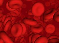
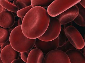
­­The cellular portion of blood contains red blood cells (RBCs), white blood cells (WBCs) and platelets. The RBCs carry oxygen from the lungs; the WBCs help to fight infection; and platelets are parts of cells that the body uses for clotting. All blood cells are produced in the bone marrow. As children, most of our bones produce blood. As we age this gradually diminishes to just the bones of the spine (vertebrae), breastbone (sternum), ribs, pelvis and small parts of the upper arm and leg. Bone marrow that actively produces blood cells is called red marrow, and bone marrow that no longer produces blood cells is called yellow marrow. The process by which the body produces blood is called hematopoiesis. All blood cells (RBCs, WBCs and platelets) come from the same type of cell, called the pluripotential hematopoietic stem cell. This group of cells has the potential to form any of the different types of blood cells and also to reproduce itself. This cell then forms committed stem cells that will form specific types of blood cells.

We'll learn more about red blood cells in detail next.

Advertisement

Red Blood Cells

red blood cells
Photo courtesy Garrigan.Net
Microscopic image of red blood cells

During formation, the RBC eventually loses its nucleus and leaves the bone marrow as a reticulocyte. At this point, the reticulocyte contains some remnants of organelles. Eventually these organelles leave the cell and a mature erythrocyte is formed. RBCs last an average of 120 days in the bloodstream. When RBCs age, they are removed by macrophages in the liver and spleen.

A hormone called erythropoietin and low oxygen levels regulate the production of RBCs. Any factor that decreases the oxygen level in the body, such as lung disease or anemia (low number of RBCs), increases the level of erythropoietin in the body. Erythropoietin then stimulates production of RBCs by stimulating the stem cells to produce more RBCs and increasing how quickly they mature. Ninety percent of erythropoietin is made in the kidneys. When both kidneys are removed, or when kidney failure is present, that person becomes anemic due to lack of erythropoietin. Iron, vitamin B-12 and folate are essential in the production of RBCs.

Red blood cells (RBCs) are by far the most abundant cells in the blood. RBCs give blood its characteristic red color. In men, there are an average of 5,200,000 RBCs per cubic millimeter (microliter), and in women there are an average of 4,600,000 RBCs per cubic millimeter. RBCs account for approximately 40 to 45 percent of the blood. This percentage of blood made up of RBCs is a frequently measured number and is called the hematocrit. The ratio of cells in normal blood is 600 RBCs for each white blood cell and 40 platelets.

There are several things about RBCs that make them unusual:

  • An RBC has a strange shape -- a biconcave disc that is round and flat, sort of like a shallow bowl.
  • An RBC has no nucleus. The nucleus is extruded from the cell as it matures.
  • An RBC can change shape to an amazing extent, without breaking, as it squeezes single file through the capillaries. (Capillaries are minute blood vessels through which oxygen, nutrients and waste products are exchanged throughout the body.)
  • An RBC contains hemoglobin, a molecule specially designed to hold oxygen and carry it to cells that need it.

The primary function of red blood cells is to transport oxygen from the lungs to the cells of the body. RBCs contain a protein called hemoglobin that actually carries the oxygen.

In the capillaries, the oxygen is released to be used by the cells of the body. Ninety-seven percent of the oxygen that is carried by the blood from the lungs is carried by hemoglobin; the other three percent is dissolved in the plasma. Hemoglobin allows the blood to transport 30 to 100 times more oxygen than could be dissolved in the plasma alone.

Hemoglobin combines loosely with oxygen in the lungs, where the oxygen level is high, and then easily releases it in the capillaries, where the oxygen level is low. Each molecule of hemoglobin contains four iron atoms, and each iron atom can bind with one molecule of oxygen (which contains two oxygen atoms, called O2) for a total of four oxygen molecules (4 * O2) or eight atoms of oxygen for each molecule of hemoglobin. The iron in hemoglobin gives blood its red color.

Thirty-three percent of an RBC is hemoglobin. The normal concentration of hemoglobin in blood is 15.5 grams per deciliter of blood in men, and 14 grams per deciliter of blood in women. (A deciliter is 100 milliliters, or one-tenth of a liter.)

Besides carrying oxygen to the cells of the body, the RBCs help to remove carbon dioxide (CO2) from the body. Carbon dioxide is formed in the cells as a byproduct of many chemical reactions. It enters the blood in the capillaries and is brought back to the lungs and released there and then exhaled as we breathe. RBCs contain an enzyme called carbonic anhydrase which helps the reaction of carbon dioxide (CO2) and water (H2O) to occur 5,000 times faster. Carbonic acid is formed, which then separates into hydrogen ions and bicarbonate ions:

Carbonic Anhydrase

CO2 + H2O ===> H2CO3 + H+ + HCO3-

carbon dioxide + water ==> carbonic acid + hydrogen ion + bicarbonate ion

The hydrogen ions then combine with hemoglobin and the bicarbonate ions go into the plasma. Seventy percent of the CO2 is removed in this way. Seven percent of the CO2 is dissolved in the plasma. The remaining 23 percent of the CO2 combines directly with hemoglobin and then is released into the lungs.

In the next section, we'll learn about the different types of white blood cells.

Advertisement

White Blood Cells

White blood cells (WBCs), or leukocytes, are a part of the immune system and help our bodies fight infection. They circulate in the blood so that they can be transported to an area where an infection has developed. In a normal adult body there are 4,000 to 10,000 (average 7,000) WBCs per microliter of blood. When the number of WBCs in your blood increases, this is a sign of an infection somewhere in your body.

Here are the six main types of WBCs and the average percentage of each type in the blood:

  • Neutrophils - 58 percent
  • Eosinophils - 2 percent
  • Basophils - 1 percent
  • Bands - 3 percent
  • Monocytes - 4 percent
  • Lymphocytes - 4 percent

Most WBCs (neutrophils, eosinophils, basophils and monocytes) are formed in the bone marrow. Neutrophils, eosinophils and basophils are also called granulocytes because they have granules in their cells that contain digestive enzymes. Basophils have purple granules, eosinophils have orange-red granules and neutrophils have a faint blue-pink color. When a granulocyte is released into the blood, it stays there for an average of four to eight hours and then goes into the tissues of the body, where it lasts for an average of four to five days. During a severe infection, these times are often shorter.

Neutrophils are the one of the body's main defenses against bacteria. They kill bacteria by actually ingesting them (this is called phagocytosis). Neutrophils can phagocytize five to 20 bacteria in their lifetime. Neutrophils have a multi-lobed, segmented or polymorphonuclear nucleus and so are also called PMNs, polys or segs. Bands are immature neutrophils that are seen in the blood. When a bacterial infection is present, an increase of neutrophils and bands are seen.

Eosinophils kill parasites and have a role in allergic reactions.

Basophils are not well understood, but they function in allergic reactions. They release histamine (which causes blood vessels to leak and attracts WBCs) and heparin (which prevents clotting in the infected area so that the WBCs can reach the bacteria).

Monocytes enter the tissue, where they become larger and turn into macrophages. There they can phagocytize bacteria (up to 100 in their lifetime) throughout the body. These cells also destroy old, damaged and dead cells in the body. Macrophages are found in the liver, spleen, lungs, lymph nodes, skin and intestine. The system of macrophages scattered throughout the body is called the reticuloendothelial system. Monocytes stay in the blood for an average of 10 to 20 hours and then go into the tissues, where they become tissue macrophages and can live for months to years.

Neutrophils and monocytes use several mechanisms to get to and kill invading organisms. They can squeeze through openings in blood vessels by a process called diapedesis. They move around using ameboid motion. They are attracted to certain chemicals produced by the immune system or by bacteria and migrate toward areas of higher concentrations of these chemicals. This is called chemotaxis. They kill bacteria by a process called phagocytosis, in which they completely surround the bacteria and digest them with digestive enzymes.

In the next section, we'll take a closer look at lymphocytes and platelets.

Advertisement

Lymphocytes and Platelets

Lymphocytes are complex cells that direct the body's immune system. T lymphocytes start in the bone marrow from pluripotent hematopoietic stem cells, then travel to and mature in the thymus gland. The thymus is located in the chest between the heart and sternum (breastbone). B lymphocytes mature in the bone marrow.

T lymphocytes (T cells) are responsible for cell-mediated immunity. B lymphocytes are responsible for humoral immunity (antibody production). Seventy-five percent of lymphocytes are T cells. Lymphocytes are different from the other WBCs because they can recognize and have a memory of invading bacteria and viruses. Lymphocytes continually pass back and forth between lymph tissue, lymph fluid and blood. When they are present in the blood, they stay for several hours. Lymphocytes can live for weeks, months or years.

There are many types of T cells that have specific functions, including:

  • Helper T cells - Helper T cells have proteins on their cell membranes called CD4. Helper T cells direct the rest of the immune system by releasing cytokines. Cytokines stimulate B cells to form plasma cells, which form antibodies, stimulate the production of cytotoxic T cells and suppressor T cells and activate macrophages. Helper T cells are the cells the AIDS virus attacks -- you can imagine that destroying the cells that direct the immune system has a devastating effect.
  • Cytotoxic T cells - Cytotoxic T cells release chemicals that break open and kill invading organisms.
  • Memory T cells - Memory T cells remain afterwards to help the immune system respond more quickly if the same organism is encountered again.
  • Suppressor T cells - Suppressor T cells suppress the immune response so that it does not get out of control and destroy normal cells once the immune response is no longer needed.

B cells become plasma cells when exposed to an invading organism or when activated by helper T cells. B cells produce large numbers of antibodies (also called immunoglobulins or gamma globulins). There are five types of immunogloulins (abbreviated Ig): IgG, IgM, IgE, IgA and IgD. These are Y-shaped molecules that have a variable segment that is a binding site for only one specific antigen. These bind to antigens, which causes them to clump, be neutralized or break open. They also activate the complement system.

The complement system is a series of enzymes that help or complement antibodies and other components of the immune system to destroy the invading antigen by attracting and activating neutrophils and macrophages, neutralizing viruses and causing invading organisms to break open. Memory B cells also remain for prolonged periods, and if the same antigen is encountered it causes a more rapid response in producing antibodies.

Platelets (thrombocytes) help blood to clot by forming something called a platelet plug. The other way that blood clots is through coagulation factors. Platelets also help to promote other blood clotting mechanisms. There are approximately 150,000 to 400,000 platelets in each microliter of blood (average is 250,000).

Platelets are formed in the bone marrow from very large cells called megakaryocytes, which break up into fragments -- these cellular fragments are platelets. They do not have a nucleus and do not reproduce. Instead, megakaryocytes produce more platelets when necessary. Platelets generally last for an average of 10 days.

Platelets contain many chemicals that assist clotting. These include:

  • Actin and myosin, to help them contract
  • Chemicals that help the coagulation process to begin
  • Chemicals that attract other platelets
  • Chemicals that stimulate blood vessel repair
  • Chemicals that stabilize a blood clot

Advertisement

Plasma

Plasma is a clear, yellowish fluid (the color of straw). Plasma can sometimes appear milky after a very fatty meal or when people have a high level of lipids in their blood. Plasma is 90-percent water. The other 10 percent dissolved in plasma is essential for life. These dissolved substances are circulated throughout the body and diffuse into tissues and cells where they are needed. They diffuse from areas of high concentration to areas of lower concentration. The greater the difference in concentration, the greater the amount of material that diffuses. Waste materials flow in the opposite direction, from where they are created in the cells into the bloodstream, where they are removed either in the kidneys or lungs.

Hydrostatic pressure (blood pressure) pushes fluid out of blood vessels. Balancing this is something called oncotic pressure (caused by proteins dissolved in blood), which tends to keep fluid inside the blood vessels.

Proteins make up a large part of the 10 percent of material dissolved in plasma and are responsible for oncotic pressure. Protein molecules are much larger than water molecules and tend to stay in blood vessels. They have more difficulty fitting through the pores in capillaries, and therefore have a higher concentration in blood vessels. Proteins tend to attract water to keep their relative concentration in blood vessels more in line with fluid outside the blood vessels. This is one of the ways the body maintains a constant volume of blood.

Plasma contains 6.5 to 8.0 grams of protein per deciliter of blood. The main proteins in plasma are albumin (60 percent), globulins (alpha-1, alpha-2, beta, and gamma globulins (immunoglobulins)), and clotting proteins (especially fibrinogen). These proteins function to maintain oncotic pressure (especially albumin) and transport substances such as lipids, hormones, medications, vitamins, and other nutrients. These proteins are also part of the immune system (immunoglobulins), help blood to clot (clotting factors), maintain pH balance, and are enzymes involved in chemical reactions throughout the body.

Electrolytes are another large category of substances dissolved in plasma. They include:

  • Sodium (Na+)
  • Potassium (K+)
  • Chloride (Cl-)
  • Bicarbonate (HCO3-)
  • Calcium (Ca+2)
  • Magnesium (Mg+2)

These chemicals are absolutely essential in many bodily functions including fluid balance, nerve conduction, muscle contraction (including the heart), blood clotting and pH balance.

Other materials dissolved in plasma are carbohydrates (glucose), cholesterol, hormones and vitamins. Cholesterol is normally transported attached to lipoproteins such as low-density lipoproteins (LDLs) and high-density lipoproteins (HDLs). For more information on cholesterol, read How Cholesterol Works.

When plasma is allowed to clot, the fluid left behind is called serum. When blood is collected from a patient it is allowed to clot in a test tube, where the cells and clotting factors fall to the bottom and the serum is left on top. Serum is tested for all the numerous items discussed above to determine if any abnormalities exist.

Advertisement

Blood Types

There are four major blood types: A, B, AB, and 0. The blood types are determined by proteins called antigens (also called agglutinogens) on the surface of the RBC.

U.S. Blood Type Distribution
According to the American Association of Blood Banking, these are the percentages of different blood types in the U.S. population:
  • A+: 34 percent
  • A-: 6 percent
  • B+: 9 percent
  • B-: 2 percent
  • AB+: 3 percent
  • AB-: 1 percent
  • O+: 38 percent
  • O-: 7 percent

There are two antigens, A and B. If you have the A antigen on the RBC, then you have type A blood. When B antigen is present, you have type B blood. When both A and B antigens are present, you have type AB blood. When neither are present, you have type O blood.

When an antigen is present on the RBC, then the opposite antibody (also called agglutinin) is present in the plasma. For instance, type A blood has anti-type-B antibodies. Type B blood has anti-type-A antibodies. Type AB blood has no antibodies in the plasma, and type O blood has both anti-type-A and anti-type-B antibodies in the plasma. These antibodies are not present at birth but are formed spontaneously during infancy and last throughout life.

In addition to the ABO blood group system, there is an Rh blood group system. There are many Rh antigens that can be present on the surface of the RBC. The D antigen is the most common Rh antigen. If the D antigen is present, then that blood is Rh+. If the D antigen is missing, then the blood is Rh-. In the United States, 85 percent of the population is Rh+ and 15 percent is Rh-. Unlike in the ABO system, the corresponding antibody to the Rh antigen does not develop spontaneously but only when the Rh- person is exposed to Rh antigen by blood transfusion or during pregnancy. When an Rh- mother is pregnant with an Rh+ fetus, then the mother forms antibodies that can travel through the placenta and cause a disease called hemolytic disease of the newborn (HDN), or erythroblastosis fetalis.

Advertisement

Donating Blood

blood supplies
Red Cross blood supplies sent to the Gulf during the Gulf War

A unit of blood is 1 pint (450 milliliters) and is mixed with chemicals (CPD) to prevent clotting. Each year, approximately 12 million to 14 million units of blood are donated in the United States. Generally, a blood donor must be at least 17 years old, be healthy, and weigh over 110 pounds.

Prior to donating blood, the donor is given an information pamphlet to read. A health history is taken to ensure that the donor has not been exposed to diseases that can be transmitted by blood, and to determine if donating blood is safe for that person's own health. The donor's temperature, pulse, blood pressure and weight are obtained. A few drops of blood are obtained to make sure the donor is not anemic. It usually takes less than 10 minutes for the blood to be removed once the needle has been placed. Sterile, single-use equipment is used so there is no danger of infection to the donor. Donors should drink extra fluids and avoid exercise that day. Blood can be donated every eight weeks.

Autologous blood donation is the donation of blood for one's own use, usually prior to surgery. Apheresis is the procedure in which only a specific component of a donor's blood is removed (usually platelets, plasma or leukocytes). In this way, more of that specific component can be removed than can be derived from one unit of blood.

Each unit of blood can be separated into several components so that each component can be given to someone with a need for that specific one. Therefore, a single unit of blood can help many people. These components include:

  • Packed RBCs
  • Fresh frozen plasma
  • Platelets
  • WBCs
  • Albumin
  • Immunoglobulins
  • Cryoprecipitate anti-hemolytic factor
  • Factor VIII concentrate
  • Factor IX concentrate

Let's look at each of these blood components in more detail.

Red blood cells (packed RBCs)

Plasma (fresh frozen plasma), once thawed, is transfused to treat bleeding disorders when many clotting factors are missing. This occurs in liver failure, when too much of a blood thinner called Coumadin has been given, or when severe bleeding and massive transfusions result in low levels of clotting factors.

Platelets are transfused in people with low platelet count (thrombocytopenia) or abnormally functioning platelets. Each unit of platelets raises the platelet count by approximately 5,000 platelets per microliter of blood.

Albumin makes up 60 percent of the protein in plasma, is produced in the liver and is used when blood volume needs to be increased and fluids have not worked, as in cases of severe bleeding, liver failure and severe burns.

Immunoglobulins are given to persons who have been exposed to a certain disease such as rabies, tetanus or hepatitis to help prevent that disease.

Factor VIII concentrate and cryoprecipitate are used in hemophilia A (classic hemophilia) since this is caused by a factor VIII deficiency.

Factor IX concentrate is used in hemophilia B ("Christmas disease"), which is caused by a deficiency of clotting factor IX.

Advertisement

Ensuring a Safe Blood Supply

There are many tests that are performed on blood to ensure its safety. These tests include checking for:
  • Hepatitis B surface antigen
  • Hepatitis B core antibody
  • Hepatitis C antibody
  • HIV-1, HIV-2 antibodies
  • HIV-1 p24 antigen
  • HTLV-1, HTLV-2 antibodies
  • Syphilis

If any of these tests are positive, the blood is discarded. As of 1996, the risk of getting HIV from a single blood transfusion was 1 in 676,000 units of blood, the risk of developing Hepatitis B was 1 in 66,000 units and the risk of getting Hepatitis C was 1 in 100,000 units. However, newer testing may decrease the risk of Hepatitis C to between 1 in 500,000 and 1 in 1,000,000.

When blood is transfused into a patient, the blood type must be determined so that a transfusion reaction does not occur.

A reaction occurs when the antigens on the RBCs of the donor blood react with the antibodies present in the recipient’s plasma. In other words, if donor blood of type A (contains A antigens) is given to someone with type B blood (they have anti-type A antibodies in their blood), then a transfusion reaction will occur.

The opposite does not occur. It is unusual for the antibodies in the plasma of the donated blood to react to the antigens on the recipients RBCs because very little plasma is transfused and it gets diluted to a level too low to cause a reaction.

When a transfusion reaction occurs, an antibody attaches to antigens on several RBCs. This causes them to clump together and plug up blood vessels. Then they are destroyed by the body (called hemolysis), releasing hemoglobin from the RBCs into the blood. Hemoglobin is broken down into bilirubin, which can cause jaundice. These events occur in hemolytic disease of the newborn (mentioned previously).

When an emergency blood transfusion is necessary and the recipient's blood type is unknown, anyone can get type O- blood transfused since type O- blood has no antigen on its surface that could react with antibodies in the recipient’s plasma. Therefore, someone with type O- blood is called a universal donor. Someone with type AB blood is called a universal recipient because they have no antibodies that could react with donated blood.

For more information on blood and related topics, check out the links on the next page.

Advertisement

How Blood Works FAQ

What is a normal platelet count?
A normal platelet count ranges from 150,000 to 450,000 platelets per microliter of blood. More than 450,000 indicates a condition called thrombocytosis while having fewer than 150,000 is known as thrombocytopenia.
What is the function of red blood cells?
The primary function of red blood cells is to transport oxygen from the lungs to the rest of the body. They also help to remove carbon dioxide (CO2) from the body, transporting it in the blood through the capillaries back to the lungs, releasing it there to be exhaled.
What are the two components of blood?
Blood is a mixture of two components: cells and plasma. The cellular portion of blood contains red blood cells, white blood cells, and platelets while the plasma is the liquid portion. Of the five liters (5.3 quarts) of blood in an adult human body, approximately 2.75 to 3 liters of blood is plasma and the rest is cellular.
What are the blood types?
The eight most common blood types are A+, A-, B+, B-, O+, O-, AB+, AB-.
What is the function of platelets?
Platelets help blood to clot by forming something called a platelet plug. When they recognize damaged blood vessels in an area of the body, they bind together.

Advertisement

Loading...