Lasik Surgery

By: Jeff Tyson
LASIK surgery corrects vision with a laser in seconds. Pictured above is the Excimer laser used in LASIK surgery.
HowStuffWorks

One of the most common physical ailments that people suffer from is poor vision. The eye is a complicated organ that requires a very exact arrangement of components to function properly. If even one of these components is not precisely the correct shape, then light that falls on the eye will not be focused correctly.

For centuries, people have relied on external lenses to alter the angle of the light entering the eye. Whether in glasses or contact lenses, these appliances have proven invaluable in the correction of poor vision. While external lenses will remain popular for the foreseeable future, advances in technology have made it possible for surgeons to alter the shape of the eye itself.

Advertisement

There are several types of vision correction surgery. One of the most popular is LASIK, which stands for laser-assisted in-situ keratomileusis. In this article, you will find out exactly what happens during a LASIK procedure as you follow this author through his own eye surgery. You will learn what LASIK is, what is involved in the surgery, what equipment is used and how to know if you're a candidate for LASIK. You will also learn what the other forms of eye surgery are and how they differ from each other.

Before we talk about laser eye surgery, let's look at how your eye works.

Advertisement

Eye to Eye

The parts of the eye

In its simplest sense, your eye is like a camera. Your eye has:

  • A variable opening called the pupil
  • A lens system, which includes the transparent covering called the cornea and a spherical lens
  • A reusable "film" called the retina
  • Various sets of muscles (The muscles control the size of the opening, the shape of the lens system and the movements of the eye.)

On the back of your eye is a complex layer of cells known as the retina. The retina reacts to light and conveys that information to the brain. The brain, in turn, translates all that activity into an image. Because the eye is a sphere, the surface of the retina is curved.

Advertisement

In the retina, sensory cells called rods and cones change the photons of light into electrical signals, which are then transmitted to and interpreted by the brain. The ability to focus the light on the retina depends on the shapes of the cornea and the lens, which are controlled by their inherent shapes, their stretchiness or elasticity, the shape of the eyeball and sets of attached muscles. So, when you look at something, muscles attached to the lens must contract and relax to change the shape of the lens system and keep the object focused on the retina, even when your eyes move; this is a complex set of muscle movements that is controlled automatically by your nervous system.

When you look at something, three things must happen:

  • The image must be reduced in size to fit onto the retina.
  • The scattered light must come together -- that is, it must focus -- at the surface of the retina.
  • The image must be curved to match the curve of the retina.

As shown below, light passes through the cornea and pupil, is bent (refracted) by the lens, and comes to a point (focus) on the retina, where the image is formed.

Light enters the eye and an image is focused on the retina.

To do all of that, the eye has a lens between the retina and the pupil (the "peep hole" in the center of your eye that allows light into the back of the eye) and a transparent covering, or cornea (the front window). The lens and the cornea work together to focus the image onto the retina.

Advertisement

Out of Focus

If you are nearsighted, the image comes into focus before it hits your retina.

Most vision problems occur when the eye cannot focus the image onto the retina. Here are a few of the most common problems:

  • Myopia (nearsightedness)
  • Hyperopia (farsightedness)
  • Astigmatism
  • Presbyopia

In nearsightedness (myopia), the light from distant objects gets focused in front of the retina rather than on it. Myopia happens usually when the eyeball is too long; however, it is sometimes caused by too much focusing power in the lens system. The result is that the person can see close-up objects clearly, but distant objects are blurry.

Advertisement

In farsightedness (hyperopia), the light gets focused in back of the retina rather than on it. Hyperopia usually happens when the eyeball is too short or when the focusing power of the lens system is too weak. The result is that a person can see distant objects clearly, but close-up objects are blurry.

If you are farsighted, the image doesn't come into focus before it hits your retina.

In astigmatism, the shape of the cornea or the lens is distorted so that the light comes into two focal points. Imagine that the lens is egg-shaped instead of spherical, and that light coming over the top and bottom edges is brought to a different focal point than light coming over the right and left sides.

In presbyopia, the cornea and lens of the eye become less stretchy, and therefore cannot change shape as readily to bring light to a focus on the retina; this happens naturally as we grow older and is usually observed when people reach their 40s. If you have presbyopia, you have trouble focusing light from near objects on the retina. To correct this problem, you might get a pair of bifocal lenses to replace your existing glasses. If you don't already wear corrective lenses, you may be able to simply use reading glasses.

LASIK is very effective in treating myopia and, in many cases, can correct vision problems resulting from astigmatism and hyperopia as well. However, presbyopia is not easily corrected through the use of laser eye surgery.

Let's take a look at exactly what LASIK is and you'll understand why it works so well for myopia.

Advertisement

What is LASIK?

To keep your hands occupied and give you something to hang onto, most ophthalmologists provide a stuffed toy for the patient to hold during surgery. Here, Lisa Wolf clings to a stuffed frog as the laser reshapes her cornea.

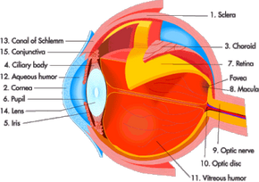
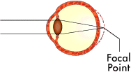
The basis for all laser eye surgery is to reshape the cornea so that it changes the focal point of the eye. Ideally, the focal point is changed so that it focuses perfectly on the retina, just like a normal eye.

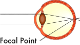
As stated in the previous section, myopia (nearsightedness) usually results from the eye being too long. The cornea has a more pronounced curve than a normal eye. Laser eye surgery is great for myopia because it is relatively easy to remove a little of the cornea to flatten out the curve.

Advertisement

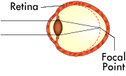
Hyperopia (farsightedness) normally means the eye is too short, which means that the cornea needs to curve more to properly focus the light on the retina. Although more intensive than correcting myopia, laser eye surgery can treat hyperopia by reshaping the cornea to make it rounder.

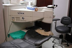
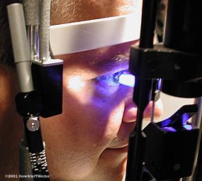
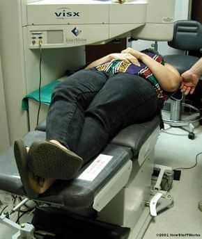
Laser eye surgery works by pulsing a tightly-focused beam of light (laser) onto the surface of the eye. Upon contact with the surface of the cornea, the laser vaporizes a microscopic portion of the cornea (more on this later). By controlling the size, position and number of laser pulses, the surgeon can precisely control how much of the cornea is removed.

LASIK combines the best features of ALK and PRK (see above). Like ALK, LASIK uses a microkeratome to create a "flap" of the outer corneal tissue that can be folded out of the way and then replaced. Once the flap is folded out of the way, LASIK uses the same Excimer laser used in PRK to reshape the underlying corneal tissue. Then the flap is replaced over the reshaped area and conforms to the new shape.

The great thing about the cornea is how quickly it heals. As soon as that flap is replaced, it begins to naturally seal itself to the rest of the cornea. This approach greatly speeds the overall healing process when compared to PRK, which leaves the reshaped area open.

Of course, there are potential problems with LASIK. The three most common problems are:

  • Undercorrection - Not enough tissue is removed during the procedure.
  • Overcorrection - Too much tissue is removed during the procedure.
  • Wrinkling - The corneal flap has a small fold or wrinkle in it when it is replaced, causing a small blurry area in your vision.

Under most circumstances, each of these problems is easily corrected with a second surgical procedure. If the undercorrection or overcorrection is very slight, the surgeon will most likely advise the patient not to attempt to refine his or her vision any further. In fact, many recipients of laser eye surgery never achieve normal vision but are able to reduce their corrective-lens prescription significantly.

In addition to the more common problems listed above, there is a potential for other side effects such as blurred vision, halos around lights, increased light sensitivity and even double vision. There is also the chance that damage or scarring can happen to the cornea, resulting in a partial or complete loss of vision.

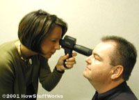
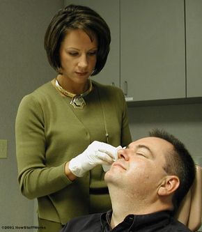
These other problems occur only rarely when you're dealing with reputable ophthalmologists operating on patients who meet the parameters of an ideal candidate. We'll talk more about what makes an ideal candidate later.

For now, let's take a closer look at the laser used in eye surgery.

Advertisement

The Excimer Laser

The laser used in my LASIK surgery is the VISX Star S3, with all of the available upgrades. The VISX Star S3 operates at 190 nanometers and the laser can adjust the treatment zone depending on pupil size (6, 6.5, 8 mm pupils). It is able to treat both nearsightedness (with/without astigmatism) and farsightedness (with/without astigmatism). This laser can also be used for therapeutic treatments of corneal scarring.

The development of the Excimer laser is the key element that has made laser eye surgery possible. Created by IBM, Excimer lasers (the name is derived from the terms excited and dimers) use reactive gases, such as chlorine and fluorine, mixed with inert gases such as argon, krypton or xenon. When electrically stimulated, a pseudo molecule (dimer) is produced that, when lased, produces light in the ultraviolet range. (See How Lasers Work for detailed information about lasers.)

The Excimer laser is a cool laser, which means that it does not heat up the surrounding air or surfaces. Instead, a very tightly-focused beam of ultraviolet light is emitted. The ultraviolet light is absorbed by the upper layer of the surface that it contacts. The sheer amount of ultraviolet light is too much for most organic materials (such as the cornea of the eye) to absorb, resulting in the breakdown of the molecular bonds of the material.

Advertisement

The ultraviolet beam of light only penetrates a microscopic amount, less than a nanometer (a billionth of a meter), into the surface of the cornea. The heat created from the energy released by the laser is dissipated along with this microscopic layer of the cornea. This process is known as photoablation.

The Excimer laser is incredibly precise. It has the ability to focus a beam as small as 0.25 microns. Considering that a typical human hair is 50 microns in diameter, that means that the Excimer laser is capable of removing 0.5 percent of a human hair's width at a time!

Bryan Lemon, Laser Engineer, makes adjustments to the Excimer laser before the surgery.

The operation of the Excimer laser is a complicated and delicate process. In fact, a dedicated technician is used just to set up and operate the machine in conjunction with the ophthalmologist performing the surgery.

Advertisement

Preoperative Visit

I gaze through the lenses of the phoreopter.

Before LASIK can be performed, you must have a thorough eye examination to ensure that you are an ideal candidate for the procedure. An ideal LASIK candidate meets the following criteria:

  • Vision correction - Your existing vision must fall within an acceptable correction range and must not have changed significantly within the last two years. The differences in vision are measured in diopters, which are degrees of prescription on a range that scales from -10.00 diopters, for severe myopia, to +4.00 diopters for severe hyperopia. A normal eye falls within the diopter range of - 0.50 to +0.50. Here are the diopter ranges that LASIK can treat: Myopia (-0.75 to -10.00) Hyperopia (+0.75 to +4.00) Astigmatism (+/- 0.75 to +/- 4.00)
  • Cornea thickness - The cornea must have a total thickness of 500 microns or greater, depending on ablation depth (how deep and how round the reshaping needs to be) and diopter range treated. The microkeratome creates a flap that is 160 microns thick and each diopter treated results in the removal of approximately 10 microns. To be considered a healthy treatment, the laser must leave about 250 to 300 microns of posterior (behind the flap) thickness after the procedure.
  • Pupil diameter - The diameter of the pupil ideally should be no more than 6.5 mm. However, advances in the laser technology can now work with diameters up to 8.5 mm.

In addition to the list above, certain conditions are considered too risky and can keep a person from being an ideal candidate:

Advertisement

  • Pregnancy - You are pregnant or attempting to become pregnant
  • Severe heart problems - Particularly if you must wear a pacemaker
  • Certain diseases - Auto-immune diseases (rheumatoid arthritis, lupus), vascular disease, eye diseases (severe glaucoma, cataracts, ocular herpes simplex), severe diabetes
  • Certain drugs - Acutane (acne), Imitrex (migraines), immune-system medications
  • Kerataconus - Condition characterized by a thinning of the cornea

It is very important that you meet the requirements for an ideal candidate. Otherwise, you greatly increase the chance for complications or poor results. Most reputable ophthalmologists will not perform the procedure on anyone who is not an ideal candidate. There are some unscrupulous doctors out there who will accept almost anyone as a LASIK patient. Many of these doctors perform LASIK for bargain rates, doing a hundred or more of the procedures a day! If you are interested in having LASIK done, be sure to research the ophthalmologist you select and check his or her success rate and patient references.

Your eyes are thoroughly checked during a scheduled pre-op (short for preoperative) visit. One thing that is done during your pre-op visit is a check to see what your current vision-correction prescription is. A device called a phoropter is used to check your prescription. Information about your existing prescription is entered into the phoropter, which combines lenses to match the prescription.

The doctor or an assistant will put together slightly different combinations of the lenses in the phoreopter while you are looking through the combined lenses at an eye chart. You will be asked which combination makes the image clearer. Then the doctor will change the combination of the one you select slightly and ask if it is better or worse. This process takes a few minutes but gives the doctor a very exact idea of what your level of vision correction is.

Another test that is performed is a manual check of the surface of the cornea for any discrepancies. Dr. Kelly uses Fluoracaine. This is a specially-formulated dye that is safe to use in the eye. A drop of Fluoracaine in the eye stains the cornea. When a blue light is shined into the eye, the Fluoracaine causes the cornea, normally clear, to glow. If there are any irregularities, a trained ophthalmologist can easily discern them.

Notice the blue glow of my eyes caused by the Fluoracaine.

Fluoracaine and the phoreopter and just a part of the pre-op process. On the next page, you will learn about the other amazing tools used to examine your eyes.

Advertisement

Focusing In

The auto-refractor calculates the approximate level of vision correction I need.

A number of cool machines are used to determine the shape and condition of your eyes. These include:

  • Auto-refractor - This machine measures the prescription in the eyes using a cone of infrared light. The infrared light is not visible to you. It is directed into your eye by the auto-refractor while you attempt to focus on an image within the machine's viewfinder. The auto-refractor changes the magnification of the image until it comes into focus for you.
  • The auto-refractor has sensors that detect the reflections from the cone of infrared light. These reflections are used to determine the size and shape of a ring at the back of the eye called the ocular fundus. This is the part of the eye directly across from the pupil's opening. By measuring the ocular fundus, the auto-refractor can determine when your eye properly focuses on the image you are staring at. The auto-refractor monitors the magnification setting and calculates the approximate level of vision correction needed. This information is then fed into the phoropter for refinement of the prescription.
  • Corneal topographer - The corneal topographer maps the clear cornea, determining its exact shape. You look into a purple, spiraled cone (sort of looks like a hypnotist's prop). Within the cone are sensors that detect infrared light.
Staring into the topographer is reminiscent of watching the psychedelic spirals from the 1960s.
It collects the light from a several hundred points across the eye. The topographer's mapping software then "connects the dots" to create an outline of the cornea's shape. This procedure is very accurate and can find anomalies in the cornea undetectable by other means.
The monitor connected to the topographer shows a color map of my corneas.
The topographer is able to precisely measure the distance and depth of each point in relation to the other points. The topographer's mapping software then "connects the dots" to create an outline of the eye's shape. This procedure is very accurate and can find anomalies in the cornea undetectable by other means.
The pupilometer used by Erika is a small, portable model.
  • Pupilometer - The other tool used during the pre-op exam is the pupilometer. This is a handheld device -- the one that Erika used for my exam looked like a ray gun from a sci-fi movie -- used to measure the exact diameter of the pupil. It does this using infrared light that is reflected back to a tiny sensor. Because the pupil reflects infrared light differently than the surrounding iris, the pupilometer's sensor can determine precisely where the pupil begins and measure the distance across it. Typically, measurements are taken with the lights on and then again with the lights off, so that the doctor can see the changes in pupil size.

Once all of the measurements are done, the ophthalmologist can determine if you are an ideal candidate. If you are considered a candidate, then you are given some information about the procedure and an opportunity to decide if you wish to continue.

Advertisement

If you decide to continue, then you must schedule an appointment for the surgery. You need to have someone accompany you since you will not be able to drive yourself home afterwards.

Advertisement

Getting Prepped

Cleaning the eyes and surrounding area

On the day of the surgery, I met with Erika Britt before the operation was scheduled to begin. Erika had me sign an informed consent. This is a document that states that you are aware of the risks involved with laser eye surgery and agree that you willingly assume those risks. Erika also went over the procedure with me so that I would know what to expect. Then she offered me a Valium to lessen the anxiety and nervousness.

After the briefing, I was led to a waiting area. When it was time for my procedure, an assistant took me into a preparation room and had me sit down while she put Alcaine in my eyes. Alcaine is a topical anesthetic that is safe to use in the eyes. The drops of Alcaine have a numbing effect that lessens the chance of any discomfort.

Advertisement

Sterilizing all the tools is an important part of the process.

I was then taken to the operating room. I laid down on a cushioned platform that was then rotated until my head was under the laser. Bryan Lemon, the laser engineer, instructed me to move my head until I was in the correct alignment. During the procedure, you cannot close your eyes. To keep this from happening, Melissa Wood, the LASIK technician that works with Dr. Kelly, used a couple of items.

  • Special tape that holds the eyelids open
  • An eyelid speculum, which is a small metal device that pushes apart the area just above and below each eye

And then the surgery...

Advertisement

The Surgery

Lisa Wolf is preparing for the actual surgery. Like me, Lisa is nearsighted, with uncorrected vision of 20/400 in both eyes.

Once the eyes were prevented from closing, a shield was placed over my right eye. The laser was calibrated over the left eye. After the calibration was completed, Dr. Kelly made two tiny marks on my cornea to indicate proper alignment of the corneal flap when the flap is replaced. The corneal flap is created using a device called a microkeratome. Dr. Kelly used a small suction ring to hold my eye steady and quickly did the cut with the microkeratome. The microkeratome fitted onto the suction ring and sliced through the top part of the cornea to create a small flap that could be folded out of the way and then put back in place after the laser was done.

As soon as Dr. Kelly had folded the corneal flap out of the way, I was told to focus on the red light above me. Then the laser was activated and the procedure carried out while I watched the flashing red light.

Advertisement

Tools of the trade

The laser was pulsed for 17 seconds. Although it was a very brief period, it seems much longer when you are lying there and trying not to move. Once the laser was done, Dr. Kelly replaced the corneal flap and administered a small amount of antibiotic to the edge of the flap. The cool thing about the cornea is that it begins to heal and rebond immediately, so there is typically no need for stitches or any other agent to hold the flap in place.

The procedure is quick and painless. Lisa told me several weeks after her surgery that her vision had stabilized at 20/20.

As soon as the left eye was done, the shield was removed from my right eye and placed over the left one. Then the surgery was repeated for the right eye. The entire procedure happened very quickly, taking less than 15 minutes from the moment that I entered the operating room until I was done. There was no pain at all. However, there was a certain amount of discomfort. This was mainly due to the fact that someone else was touching my eyes, leaving me with a nearly uncontrollable urge to pull away or close my eyes. It was a good thing that my eyes were taped open.

Advertisement

Post-Op

After the surgery, I was taken back to the preparation room. The most incredible thing to me was that I could already see! My vision was a little blurry, but I could already tell that it was much improved from what it had been without glasses. Erika gave me a set of eye shields.

These are two silvery plastic ovals attached to an elastic band. Each oval has dozens of tiny holes in it. This allows you to see just enough to get around, but prevents you from touching your eyes at all. I was instructed to wear those for the remainder of that day and to sleep in them that night. I was also given three items to put into my eyes:

Advertisement

  • Rewetting drops like the ones used by people who wear contacts to keep the eyes from drying out
  • Antibiotic drops that reduce the chance of infection
Antibiotic drops help the eyes fight off any potential infection.
  • Moisturizing gel that is placed on the inside of your bottom eyelid just before you go to bed. It provides moisture through the night like the rewetting drops do during the day.

Advertisement

The Day After

The change in my vision is phenomenal.

The most uncomfortable thing to me was wearing the eye shields. It was like wearing a Lone Ranger mask all day and all night. But all of the discomfort was forgotten the next morning when I woke up and looked around. I could see! I looked over at the digital clock on the night stand and could easily make out the numbers. I turned on the television and enjoyed watching the early morning news. Most importantly, my eyes did not hurt at all. No itchiness, swelling or even unusual dryness. I still used the drops just like Erika had told me to. There was a little redness from the suction device and my eyesight was still a little blurry. Also, I was just a little bit sensitive to light. Dr. Kelly had told me to expect that blurriness and sensitivity for a couple of weeks as my eyes completely healed.

The follow-up appointments were scheduled on a progressively lengthening basis:

  • One day after surgery
  • One week after surgery
  • One month after surgery
  • Three months after surgery
  • Six months after surgery
  • One year after surgery

Later in the afternoon of the day immediately after my surgery, I went back to Dr. Kelly for my first follow-up appointment. Erika gave me a vision test to see what my improvement was. She said that the average result for the first day after surgery is 20/50. My right eye was 20/25 and my left eye was 20/20, an amazing leap from the 20/400 that I had in both eyes the day before! Dr. Kelly then checked each eye to make sure that it was healing properly and gave me a clean bill of health.

My next appointment was a week after the surgery. Erika once again checked my eyesight. It had continued to improve over the week and was now 20/15, even better than normal! Erika quickly cautioned me that my vision could regress slightly over the next couple of months but should not regress beyond 20/20. Dr. Kelly once again checked my eyes and said that they were doing great.

At my one-month exam, Dr. Kelly informed me that my eyes seem to be completely healed with no side effects whatsoever. My vision has stabilized between 20/20 and 20/15. Life is good!

For more information on LASIK and related topics, check out the links on the next page.

Advertisement

Loading...