How Muscles Work

Muscles of the human body
Photo courtesy National Library of Medicine

Muscles are one of those things that most of us take completely for granted, but they are incredibly important for two key reasons:

  • Muscles are the "engine" that your body uses to propel itself. Although they work differently than a car engine or an electric motor, muscles do the same thing -- they turn energy into motion.
  • It would be impossible for you to do anything without your muscles. Absolutely everything that you conceive of with your brain is expressed as muscular motion. The only ways for you to express an idea are with the muscles of your larynx, mouth and tongue (spoken words), with the muscles of your fingers (written words or "talking with your hands") or with the skeletal muscles (body language, dancing, running, building or fighting, to name a few).

­Because muscles are so crucial to any animal, they are incredibly sophisticated. They are efficient at turning fuel into motion, they are long-lasting, they are self-healing and they are able to grow stronger with practice. They do everything from allowing you to walk to keeping your blood flowing!

Advertisement

­When most people think of "muscles," they think about the muscles that we can see. For example, most of us know about the biceps muscles in our arms. But there are three unique kinds of muscle in any mammal's body:

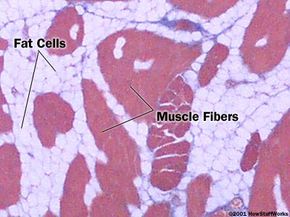
  • Skeletal muscle is the type of muscle that we can see and feel. When a body builder works out to increase muscle mass, skeletal muscle is what is being exercised. Skeletal muscles attach to the skeleton and come in pairs -- one muscle to move the bone in one direction and another to move it back the other way. These muscles usually contract voluntarily, meaning that you think about contracting them and your nervous system tells them to do so. They can do a short, single contraction (twitch) or a long, sustained contraction (tetanus).
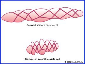
  • Smooth muscle is found in your digestive system, blood vessels, bladder, airways and, in a female, the uterus. Smooth muscle has the ability to stretch and maintain tension for long periods of time. It contracts involuntarily, meaning that you do not have to think about contracting it because your nervous system controls it automatically. For example, your stomach and intestines do their muscular thing all day long, and, for the most part, you never know what's going on in there.
  • Cardiac muscle is found only in your heart, and its big features are endurance and consistency. It can stretch in a limited way, like smooth muscle, and contract with the force of a skeletal muscle. It is a twitch muscle only and contracts involuntarily.

In this article, we will look at the different types of muscles in your body and the amazing technology that allows them to work so well.  From here on, we will focus on skeletal muscle. The basic molecular processes are the same in all three types.


Advertisement

Skeletal-muscle Basics

Cross section of a skeletal muscle (200x) showing the muscle fibers (red) and the fat cells (white)

Skeletal muscle is also called striated muscle, because when it is viewed under polarized light or stained with an indicator, you can see alternating stripes of light and dark.

Skeletal muscle has a complex structure that is essential to how it contracts. We will tease apart a skeletal muscle, starting with the largest structures and working our way to the smaller ones.

Advertisement

The basic action of any muscle is contraction. For example, when you think about moving your arm using your biceps muscle, your brain sends a signal down a nerve cell telling your biceps muscle to contract. The amount of force that the muscle creates varies -- the muscle can contract a little or a lot depending on the signal that the nerve sends. All that any muscle can do is create contraction force.

A muscle is a bundle of many cells called fibers. You can think of muscle fibers as long cylinders, and compared to other cells in your body, muscle fibers are quite big. They are from about 1 to 40 microns long and 10 to 100 microns in diameter. For comparison, a strand of hair is about 100 microns in diameter, and a typical cell in your body is about 10 microns in diameter.

A muscle fiber contains many myofibrils, which are cylinders of muscle proteins. These proteins allow a muscle cell to contract. Myofibrils contain two types of filaments that run along the long axis of the fiber, and these filaments are arranged in hexagonal patterns. There are thick and thin filaments. Each thick filament is surrounded by six thin filaments.

Thick and thin filaments are attached to another structure called the Z-disk or Z-line, which runs perpendicular to the long axis of the fiber (the myofibril that runs from one Z-line to another is called a sarcomere). Running vertically down the Z-line is a small tube called the transverse or T-tubule, which is actually part of the cell membrane that extends deep inside the fiber. Inside the fiber, stretching along the long axis between T-tubules, is a membrane system called the sarcoplasmic reticulum, which stores and releases the calcium ions that trigger muscle contraction.

Advertisement

Contracting a Muscle

During contraction, the thin filaments slide past the thick filaments, shortening the sarcomere.

The thick and thin filaments do the actual work of a muscle, and the way they do this is pretty cool. Thick filaments are made of a protein called myosin. At the molecular level, a thick filament is a shaft of myosin molecules arranged in a cylinder. Thin filaments are made of another protein called actin. The thin filaments look like two strands of pearls twisted around each other.

Advertisement

During contraction, the myosin thick filaments grab on to the actin thin filaments by forming crossbridges. The thick filaments pull the thin filaments past them, making the sarcomere shorter. In a muscle fiber, the signal for contraction is synchronized over the entire fiber so that all of the myofibrils that make up the sarcomere shorten simultaneously.

There are two structures in the grooves of each thin filament that enable the thin filaments to slide along the thick ones: a long, rod-like protein called tropomyosin and a shorter, bead-like protein complex called troponin. Troponin and tropomyosin are the molecular switches that control the interaction of actin and myosin during contraction.

While the sliding of filaments explains how the muscle shortens, it does not explain how the muscle creates the force required for shortening. To understand how this force is created, let's think about how you pull something up with a rope:

  1. Grab the rope with both hands, arms extended.
  2. Loosen your grip with one hand, let's say the left hand, and maintain your grip with the right.
  3. With your right hand holding the rope, change your right arm's shape to shorten its reach and pull the rope toward you.
  4. Grab the rope with your extended left hand and release your right hand's grip.
  5. Change your left arm's shape to shorten it and pull the rope, returning your right arm to its original extended position so it can grab the rope.
  6. Repeat steps 2 through 5, alternating arms, until you finish.

Muscles create force by cycling myosin crossbridges.

To understand how muscle creates force, let's apply the rope example.

Myosin molecules are golf-club shaped. For our example, the myosin clubhead (along with the crossbridge it forms) is your arm, and the actin filament is the rope:

  1. During contraction, the myosin molecule forms a chemical bond with an actin molecule on the thin filament (gripping the rope). This chemical bond is the crossbridge. For clarity, only one cross-bridge is shown in the figure above (focusing on one arm).
  2. Initially, the crossbridge is extended (your arm extending) with adenosine diphosphate (ADP) and inorganic phosphate (Pi) attached to the myosin.
  3. As soon as the crossbridge is formed, the myosin head bends (your arm shortening), thereby creating force and sliding the actin filament past the myosin (pulling the rope). This process is called the power stroke. During the power stroke, myosin releases the ADP and Pi.
  4. Once ADP and Pi are released, a molecule of adenosine triphosphate (ATP) binds to the myosin. When the ATP binds, the myosin releases the actin molecule (letting go of the rope).
  5. When the actin is released, the ATP molecule gets split into ADP and Pi by the myosin. The energy from the ATP resets the myosin head to its original position (re-extending your arm).
  6. The process is repeated. The actions of the myosin molecules are not synchronized -- at any given moment, some myosins are attaching to the actin filament (gripping the rope), others are creating force (pulling the rope) and others are releasing the actin filament (releasing the rope).

The contractions of all muscles are triggered by electrical impulses, whether transmitted by nerve cells, created internally (as with a pacemaker) or applied externally (as with an electrical-shock stimulus).

Advertisement

Triggering and Reversing Contraction

The coupling process leading from electrical signal (excitation) to contraction in skeletal muscle

The trigger for a muscle contraction is an electrical impulse. The electrical signal sets off a series of events that lead to crossbridge cycling between myosin and actin, which generates force. The series of events is slightly different between skeletal, smooth and cardiac muscle.

Advertisement

Let's take a look at what occurs within a skeletal muscle, from excitation to contraction to relaxation:

  1. An electrical signal (action potential) travels down a nerve cell, causing it to release a chemical message (neurotransmitter) into a small gap between the nerve cell and muscle cell. This gap is called the synapse.
  2. The neurotransmitter crosses the gap, binds to a protein (receptor) on the muscle-cell membrane and causes an action potential in the muscle cell.
  3. The action potential rapidly spreads along the muscle cell and enters the cell through the T-tubule.
  4. The action potential opens gates in the muscle's calcium store (sarcoplasmic reticulum).
  5. Calcium ions flow into the cytoplasm, which is where the actin and myosin filaments are.
  6. Calcium ions bind to troponin-tropomyosin molecules located in the grooves of the actin filaments. Normally, the rod-like tropomyosin molecule covers the sites on actin where myosin can form crossbridges.
  7. Upon binding calcium ions, troponin changes shape and slides tropomyosin out of the groove, exposing the actin-myosin binding sites.
  8. Myosin interacts with actin by cycling crossbridges, as described previously. The muscle thereby creates force, and shortens.
  9. After the action potential has passed, the calcium gates close, and calcium pumps located on the sarcoplasmic reticulum remove calcium from the cytoplasm.
  10. As the calcium gets pumped back into the sarcoplasmic reticulum, calcium ions come off the troponin.
  11. The troponin returns to its normal shape and allows tropomyosin to cover the actin-myosin binding sites on the actin filament.
  12. Because no binding sites are available now, no crossbridges can form, and the muscle relaxes.

As you can see, muscle contraction is regulated by the level of calcium ions in the cytoplasm. In skeletal muscle, calcium ions work at the level of actin (actin-regulated contraction). They move the troponin-tropomyosin complex off the binding sites, allowing actin and myosin to interact.

All of this activity requires energy. Muscles use energy in the form of ATP. The energy from ATP is used to reset the myosin crossbridge head and release the actin filament. To make ATP, the muscle does the following:

  1. Breaks down creatine phosphate, adding the phosphate to ADP to create ATP
  2. Carries out anaerobic respiration, by which glucose is broken down to lactic acid and ATP is formed
  3. Carries out aerobic respiration, by which glucose, glycogen, fats and amino acids are broken down in the presence of oxygen to produce ATP (see How Exercise Works for details).

Muscles have a mixture of two basic types of fibers: fast twitch and slow twitch. Fast-twitch fibers are capable of developing greater forces, contracting faster and have greater anaerobic capacity. In contrast, slow-twitch fibers develop force slowly, can maintain contractions longer and have higher aerobic capacity. Training can increase muscle mass, probably by changing the size and number of muscle fibers rather than the types of fibers. Some athletes also use performance-enhancing drugs, specifically anabolic steroids, to build muscle, although this practice is dangerous and is banned in most athletic competitions.

Advertisement

Cardiac and Smooth Muscle

While most of the processes are similar, there are some notable differences between the actions of skeletal, cardiac and smooth muscle.

Cardiac-muscle cells are striated, and are a lot like skeletal-muscle cells except that in cardiac muscle, the fibers are interconnected. The sarcoplasmic reticulum of cardiac-muscle cells is not as well-developed as that of skeletal-muscle cells. Cardiac-muscle contraction is actin-regulated, meaning that the calcium ions come both from the sarcoplasmic reticulum (as in skeletal muscle) and from outside the cell (as in smooth muscle). Otherwise, the chain of events that occurs in cardiac-muscle contraction is similar to that of skeletal muscle.

Advertisement

Compared to skeletal muscle, smooth-muscle cells are small. They are spindle-shaped, about 50 to 200 microns long and only 2 to 10 microns in diameter. They have no striations or sarcomeres. Instead, they have bundles of thin and thick filaments (as opposed to well-developed bands) that correspond to myofibrils. In smooth-muscle cells, intermediate filaments are interlaced through the cell much like the threads in a pair of "fish-net" stockings. The intermediate filaments anchor the thin filaments and correspond to the Z-disks of skeletal muscle. Unlike skeletal-muscle cells, smooth-muscle cells have no troponin, tropomyosin or organized sarcoplasmic reticulum.

As in skeletal-muscle cells, contraction in a smooth-muscle cell involves the forming of crossbridges and thin filaments sliding past thick filaments. However, because smooth muscle is not as organized as skeletal muscle, shortening occurs in all directions. During contraction, the smooth-muscle cell's intermediate filaments help to draw the cell up, like closing a drawstring purse.

Calcium ions regulate contraction in smooth muscle, but they do it in a slightly different way than in skeletal muscle:

  1. Calcium ions come from outside of the cell.
  2. Calcium ions bind to an enzyme complex on myosin, called calmodulin-myosin light chain kinase.
  3. The enzyme complex breaks up ATP into ADP and transfers the Pi directly to myosin.
  4. This Pi transfer activates myosin.
  5. Myosin forms crossbridges with actin (as occurs in skeletal muscle).
  6. When calcium is pumped out of the cell, the Pi gets removed from myosin by another enzyme.
  7. The myosin becomes inactive, and the muscle relaxes.

This process is called myosin-regulated contraction.

For more information on muscles and related topics, check out the links on the next page.

Advertisement

How Muscles Work FAQ

What is the strongest muscle in the human body?
There's no agreement because there are multiple ways to judge strength. Some experts believe it's the masseter (used for chewing), while others claim it's the gluteus maximus (buttocks).
What are the types of muscles?
The three types of muscle tissue are cardiac, smooth, and skeletal or striated. Cardiac muscle is found only in the heart while smooth muscle is found in the digestive system, blood vessels, bladder, airways and uterus. Skeletal muscle is the type of muscle that you can see and feel.
How many muscles and bones are in the human body?
It's estimated that there are more than 650 named skeletal muscles in your body. The adult human body contains 206 bones.
What triggers a muscle contraction?
A muscle contraction is triggered by an electrical impulse (excitation) when the brain sends a signal along the nerves to the muscle.
How do muscles move the body?
Muscles move the body by contracting and relaxing. Muscles can pull bones, but they can't push them, so they work in pairs of flexors and extensors. Fast-twitch muscle fibers can contract faster, while slow-twitch fibers can maintain contractions longer.

Advertisement

Loading...