How can someone be born with organs outside the body?

human internal organs
Bodily Organs Image Gallery A gastroschisis or omphalocele occurs when a person is born with his intestines or other internal organs outside his body.  See more bodily organ pictures.
Purestock/Getty Images

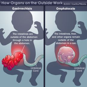
You may have read stories about babies born with a protruding liver or ropes of intestine hanging outside the abdomen. While this idea may seem far-fetched, these types of conditions occur more often than you might think. In certain forms, they occur as often as one in 5,000 births.

These birth defects can be life threatening, especially since they come with the risk of other defects, infection and complications. The good news is that we've learned a lot about these conditions over the past few decades. With this information, doctors have improved detection and treatment methods dramatically. Let's take a look at two conditions in which organs, or parts of organs, appear outside of the body: gastroschisis and omphalocele.

Advertisement

Both of these conditions happen when something goes awry during organ development. Between six and 10 weeks after conception, the baby's intestines, stomach and liver jut into the umbilical cord. By week 10, the baby's intestines usually go back into the abdomen. Throughout these early stages of development, there's a small opening in the baby's abdominal muscles, which the umbilical cord passes through on its way to the placenta. In later stages of development, these abdominal muscles grow together to close this tiny opening. When the abdominal organs don't return to the abdominal cavity and the muscles fail to close, this is called either gastroschisis or omphalocele, depending on the severity.

We'll look at gastroschisis -- the less severe condition -- on the next page.


Advertisement

Gastroschisis

illustration organs outside

Gastroschisis, meaning "belly cleft" in Greek, usually appears as a 2- to 4-centimeter opening of the abdomen on the right side of the umbilical cord [source: American Pediatric Surgical Association]. Although less severe than an omphalocele, gastroschisis is still a dangerous condition that leads to death in 15 percent of cases [source: Engber]. Gastroschisis is the most frequently occurring problem with the abdominal wall, and 40 percent of babies born with gastroschisis are undersized or born prematurely [source: American Pediatric Surgical Association].

With gastroschisis, nothing is covering the exposed intestine, causing it to come into contact with the amniotic fluid when the fetus is still in the womb. When this happens, a hard film develops around the intestine, which hinders the newborn's intestinal function, sometimes for months.

Advertisement

With such a sensitive part of the body exposed, there's a chance for dangerous complications to occur. One complication, called atresia, occurs in 10 to 15 percent of gastroschisis cases [source: American Pediatric Surgical Association]. With atresia, a portion of the intestine doesn't form at all, perhaps because blood didn't reach that area or because the intestine became twisted and died. Other possible complications include a ruptured intestine, loss of fluid and nutrients, or internal reproductive organs appearing outside the abdomen.

Doctors rely on a prenatal ultrasound to diagnose gastroschisis. Once the baby is born, doctors immediately take action to deal with the birth defect, administering antibiotics to combat infection. The baby may also be put in a sterile bowel bag that rises up to the chest and provides insulation from dangerous infections. If the abdomen is large enough to receive the protruding organs -- and in many cases it's not -- the opening may be surgically closed after birth.

In many cases, the abdomen is too small, so a device called a preformed silo is used to gradually soften the intestines and ease them back into the abdomen. Then, when the baby is a couple of weeks old, doctors perform surgery to seal the hole [source: American Pediatric Surgical Association. Barring further complications, the baby has a good chance for recovery.

Now let's take a look at a more severe birth defect involving organs on the outside of the body.

Advertisement

Omphalocele

ultrasound with doctors
Ultrasounds can help detect omphalocele before a baby is born.
Thierry Dosogne/Iconica/Getty

Like gastroschisis, an omphalocele is a birth defect in which organs normally located in the body develop outside the body because of an opening near the umbilical cord. It can range in severity from mild to catastrophic but is generally larger and more dangerous than gastroschisis. With a small-type omphalocele, only a small piece of intestine pokes through the hole in the abdomen. A large-type omphalocele occurs when several major organs, such as the liver, intestines, spleen, ovaries and testes, appear outside the body.

Like gastroschisis, an omphalocele can be diagnosed during pregnancy through ultrasound, but only after the first trimester. The causes of this defect remain unknown. Small-type omphaloceles occur in one in 5,000 births, and large-type omphaloceles are found in one in 10,000 births [source: University of Virginia Health System]. Generally, omphaloceles are more dangerous than gastroschisis because they often appear with other birth defects -- this happens in twenty-five to 40 percent of babies born with omphaloceles [source: MedlinePlus]. Babies with large-type omphaloceles may also have lung and heart problems and chromosomal abnormalities.

Advertisement

A small-type omphalocele can sometimes be repaired through surgery not long after birth. The treatment of a large-type omphalocele is more complicated. With a large-type omphalocele, the organs developing on the outside of the body are protected by a membrane, but this membrane is fragile. If it breaks, the subsequent infections almost always prove fatal. Making the situation more delicate, the newborn's abdomen is usually too small for surgeons to surgically implant the organs in the body.

To protect the membrane-covered organs, doctors often use a treatment called paint and wait [source: Hageman]. Doctors prescribe an antibiotic cream that's regularly applied to the membranous sac surrounding the organs. The membrane hardens while skin develops and begins to cover parts of the organs. Once the baby has grown and the situation is stabilized, surgeons close the abdominal cavity with the organs tucked inside.

While these birth defects can sometimes be life threatening, advancements in medicine often makes them treatable. For more information about organs developing outside of the body, birth defects and other related topics, please follow the links on the next page.

Advertisement

Lots More Information

Related Articles

More Great Links

  • Engber, David. "The Case of the Inside-Out Baby." Slate. Sept. 28, 2006. http://www.slate.com/id/2150559/
  • Hageman, B. "6-week-old Naperville baby fights the odds against her survival." Chicago Tribune. April 20, 2008. http://www.chicagotribune.com/features/lifestyle/chi-0420_baby_d_k_napr20,0,3987695.story
  • "Cloacal exstrophy." Children's Hospital Boston. http://www.childrenshospital.org/az/Site2187/mainpageS2187P0.html
  • "Gastroschisis." American Pediatric Surgical Association. 2003. http://www.eapsa.org/Gastroschisis.htm
  • "Gastroschisis." Medline Plus. National Library of Medicine and National Institute of Health. Oct. 8, 2007. http://www.nlm.nih.gov/medlineplus/ency/article/000992.htm
  • "Omphalocele." Medline Plus. National Library of Medicine and National Institute of Health. Oct. 8, 2007. http://www.nlm.nih.gov/medlineplus/ency/article/000994.htm
  • "Omphalocele." University of Virginia Health System. Jan. 28, 2008. http://www.healthsystem.virginia.edu/UVAHealth/peds_hrnewborn/omphaloc.cfm

Advertisement

Loading...