How Prenatal Testing Works

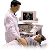
Ultrasound examination during pregnancy is one common form of prenatal testing. See more pregnancy pictures.
Photo courtesy of Philips Research

Perhaps you or someone you know is having a baby. One of the most important questions (or worries) during a pregnancy is "Is the baby healthy?" or "Is the baby developing properly?" After all, the uterus in which the baby grows is essentially a "black box" relative to the outside world. How do we know that a baby is developing properly throughout the 40 weeks of gestation?

In this article, we will examine the tests that obstetricians (the doctors who care for pregnant women during pregnancy and delivery) use to assess the development of an unborn child.

Advertisement


Advertisement

Pregnancy Involves Many Tests

Throughout the course of a pregnancy, an expecting mother will have many tests. They fall into the following categories:

  • pregnancy test - the first test
  • routine, non-invasive tests - these occur during each visit to the obstetrician's office: blood pressure, urine glucose/protein, fetal heartbeat
  • blood sampling tests - usually done once at various times during pregnancy: blood type, Rh Factor, determine iron levels, HIV, Hepatitis B, syphilis, glucose tolerance test, triple screen test
  • swab tests - usually done once at various times during pregnancy: Pap smear - to check for STDs, various bacteria (varies according to the laws of each state); Group B streptococcus screening
  • ultrasound - done at least one time during pregnancy
  • tests done under special circumstances: amniocentesis, chorionic villus sampling, fetal blood sampling or percutaneous umbilical blood sampling, fetal monitoring

Let's look at the various tests to see how they work and what they can tell us about the developing baby.

Advertisement

The Pregnancy Test

This test is usually the first test conducted when you suspect that you may be pregnant. There are a variety of home testing kits available over-the-counter and all detect a protein hormone called human chorionic gonadotropin (hCG). When an egg is fertilized, the embryo begins to produce hCG. Levels of hCG increase after conception and can be detected in the mother's urine. By 10 days after conception, hCG levels are about 25 milli-International Units (mIU).

Typically, the home test is a urine test for hCG:

Advertisement

  1. You collect a sample of urine. You would usually use the first urine in the morning, when hCG levels are the most concentrated, or wave the test wand through the urine stream.
  2. If you collected the urine, you can either dip the test wand into the cup or place a drop on the test wand.
  3. The test wands or dipsticks have a plastic coating embedded with antibodies to hCG.
  4. The test wands also have a second antibody to hCG linked with some color tag (e.g., colored latex beads, enzyme that produces a color reaction).
  5. If sufficient levels of hCG are present in the urine (more than 25 mIU), then the hCG will bind with the second antibody and cause a color reaction to occur (i.e., a positive test result).

If a positive test occurs, you generally call your doctor and a second test is performed at the office to confirm the pregnancy. The doctor may also order a blood test to determine the precise quantity of hCG present, which can be used to assess the baby's health.

Advertisement

Routine Non-Invasive Tests

These tests are done every time you visit your obstetrician and include:

  • blood pressure
  • urine glucose
  • urine protein
  • fetal heartbeat - beginning when the baby's heart is developed enough to to be heard

Blood Pressure

The increased blood volume and fetal blood circulation that occurs in pregnancy increases the demands on your cardiovascular system, especially your heart. So, your blood pressure will be measured regularly to detect any signs of high blood pressure or pregnancy-induced hypertension. About five percent of pregnant women experience pregnancy-induced hypertension starting about the 20th week of pregnancy. This condition can can lead to the following complications:

Advertisement

  • Pre-term labor
  • Separation of the placenta, leading to bleeding
  • Reduced kidney function or failure
  • Reduced blood flow to the baby, which can retard its growth and development

Pregnancy-induced hypertension, along with swelling (edema) and protein in the urine (albuminuria), comprise a condition known as preeclampsia. The cause of pre-eclampsia is unknown and the treatment is premature delivery of the baby, if possible. Sometimes, high doses of magnesium sulfate can be given to delay the symptoms until the baby can be delivered safely; no one knows why this treatment can work.

Your blood pressure will be measured with a blood pressure gauge or sphygmomanometer (read this question about blood pressure gauges for more details).

Urine Glucose

During each doctor's visit, you will be asked to pass a test strip through your urine stream or collect a sample of urine, which will be tested with a strip that measures the amount of glucose in your urine. The presence of glucose in the urine is an indication of gestational diabetes, a form of diabetes that usually develops around the 20th week of pregnancy. Gestational diabetes causes the following complications:

  • The baby grows larger than normal and develops more fat. Large babies are difficult to deliver.
  • The baby's pancreas must secrete large amounts of insulin to get rid of the excess sugar coming from the mother. After birth, when the baby is no longer receiving these high amounts of sugar from the mother, the high insulin levels can cause the baby's blood sugar to fall dangerously low (i.e., hypoglycemia).
  • Some babies from mothers with gestational diabetes have trouble breathing when they are delivered (i.e., respiratory distress).

Gestational diabetes can be treated usually by monitoring the mother's diet. However, sometimes the mother must take insulin to control her blood glucose levels. Gestational diabetes in the mother usually goes away once the baby has been delivered.

The test strip contains two enzymes (glucose oxidase and peroxidase), a chemical (orthotolidine) and a yellow dye impregnated in the paper. The reactions go like this:

  1. Glucose oxidase converts glucose into gluconic acid and hydrogen peroxide.
  2. Peroxidase reacts the hydrogen peroxidase with orthotolidine to produce a blue color.
  3. The yellow dye spreads the color change out over a wider range in proportion to the amount of glucose present.

If no glucose is present, then the test strip remains yellow. If glucose is present, then the color can vary from light green to dark blue, depending upon the concentration of glucose in the urine.

Urine Protein

The presence of protein in the urine indicates a problem in kidney function and is one of the symptoms of pre-eclampsia, as mentioned above. To detect protein in the urine, the test strip has a pH buffer (citrate buffer) and a color indicator (bromphnol blue) impregnated in the paper. At the normal pH of the paper, most of the indicator is not ionized. Proteins can bind to the nonionized form and release hydrogen ions, which changes the pH and the color of the paper. If protein is present, then the color of the paper will change from yellow to green or blue, depending upon the concentration of protein.

Fetal Heartbeat

One of the more emotional times in an early pregnancy may be the first time you hear the baby's heartbeat. The baby's heartbeat can be seen in a Doppler ultrasound as early as five to six weeks of development. By 12 to 13 weeks, your doctor can hear the heartbeat using a specialized ultrasound stethoscope or Doppler stethoscope. The Doppler stethoscope works like a regular ultrasound machine except that it does not give an image. Instead, the echoes are counted and the count is displayed on a LCD readout. If the stethoscope has a speaker, you can hear the baby's amplified heart beat.
Advertisement

Blood Sampling Tests

In the first trimester of pregnancy, you may have blood drawn to determine the following:

  • hCG levels (discussed above)
  • Blood iron content - important for hemoglobin and delivering sufficient oxygen to the growing baby
  • Blood type and Rh compatibility - assess problems between mother and baby's blood types (see Rh Blood Testing for details)
  • Presence of antibodies to viruses (e.g., HIV, hepatitis B) or presence of bacteria (e.g., syphilis).

The hCG, viral antibody, syphilis and blood type tests look for and/or quantify the amount of antibodies to these substance in the blood. The iron test directly measures the iron by burning a sample in a high temperature flame and quantifying the amount of light given off at the wavelength of light specific for iron, which is related to the amount of iron present.

Advertisement

Glucose Tolerance Test

Between week 25 to 28 gestation, you will have a glucose screening test for gestational diabetes. You will drink a soda containing high amounts of glucose, or sugar, and will have your blood drawn one hour later. Blood glucose will be measured by a glucose oxidase reaction described in the previous section. If the glucose level is high, you may be asked to take an additional glucose-tolerance test. In this test, you drink a high-glucose solution on an empty stomach and blood samples will be taken at regular intervals (usually every hour) for three hours. Blood glucose levels will be measured again. The timecourse information from a glucose tolerance test is a better indication of your body's response to a glucose load to diagnose gestational diabetes.

Triple Screen Test

This test is done in the second trimester and measures three parameters:

  • alpha-fetoprotein (AFP)
  • hCG
  • estriol

AFP is produced by the baby and makes its way into the mother's blood. Typically, AFP levels are low. However, high levels of AFP indicate that the baby's neural tube has failed to close (i.e., a neural tube defect). An ultrasound examination may then be done to confirm this finding.

At this time in development, hCG levels in combination with AFP levels can tell us whether the baby has any abnormalities in the number of chromosomes. A high level of hCG in combination with a low level of AFP suggests a chromosomal abnormality. The most common disorder of this type is Down syndrome (extra chromosome #21 -- see Human Chromosomal Abnormalities for more details). If the level of hCG is high and no fetal heartbeat is detected, then there may be a molar pregnancy. A molar pregnancy is when a piece of tissue, usually leftover from a previous pregnancy, grows rapidly, destroys the baby and forms a benign tumor. An ultrasound examination may then be done to confirm this finding.

Estriol is a hormone made by the baby's adrenal glands. The levels of estriol in the mother's blood indicate the health of the baby. If the estriol levels drop, then the baby may be threatened and need to be delivered. Low levels of estriol may also indicate Down syndrome or neural tube defect.

Advertisement

Swab Tests

At different times during pregnancy, your doctor may take swab samples (Q-tip) from your vagina or rectum. The cells on the swab will be examined under a microscope or cultured to determine various conditions or diseases.

In the first trimester, you may have a Pap smear in which cells from your cervix are examined to look for signs of cervical cancer. Later, when you are closer to delivery (around week 35), your doctor will take swabs from your vagina and rectum. These swabs will be cultured to look for Group B streptococcus bacteria. These bacteria are common in many women and are considered a normal part of an adult woman's reproductive system. For an unborn child, however, these bacteria can spread during delivery and cause several problems:

Advertisement

  • respiratory distress - problems with breathing that threaten the baby's life
  • mental retardation
  • vision problems
  • hearing loss

Group B strep infections can be treated with IV antibiotics before delivery.

Advertisement

Ultrasound

You may have one or more ultrasound tests during your pregnancy. The first test may be done within the first four weeks to date the pregnancy and determine the due date. This is done by measuring the baby's crown to rump length. Accurate charts are available to determine the age of the fetus from these measurements.

By about weeks 11 to 13, ultrasound may be used to examine various aspects of your baby's developments, such as:

Advertisement

  • Is the neural tube closed?
  • Are the internal organs developing properly?
  • Does the heart beat OK, and at what rate?

By 20 weeks, you may be able to determine the sex of the baby by observing its sex organs with ultrasound. For more details on ultrasound, see How Ultrasound Works.

Advertisement

Amniocentesis

If problems with the baby's development are detected by other means or the parents have risk factors for various genetic diseases, special tests may be required. For example, if the mother is age 34 or older, she has an increased risk of having a baby with Down syndrome. So, the parents, in consultation with the obstetrician, may elect to sample fluid or tissue from the baby to determine its genetic make-up. These samples can be obtained through three procedures:

In these tests, the doctor uses a needle or suction tube to sample fluids or the baby's tissue with the aid of ultrasound to see what he/she is doing. The tissue/fluid samples are analyzed in a laboratory to determine the number of chromosomes (karyotype) and other biochemical tests (e.g., AFP). The results from the tests can be used to make decisions about possibly terminating the pregnancy or preparing the parents to deal with any genetic defects (genetic counseling).

Advertisement

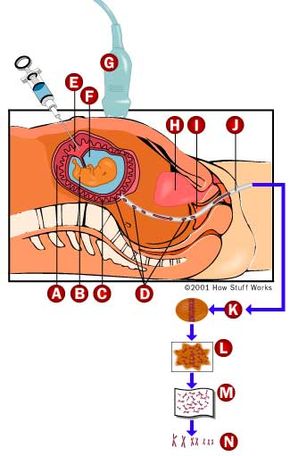
Amniocentesis is a test in which the doctor samples the fluid (amniotic fluid) that surrounds the growing baby in the uterus. Floating in the amniotic fluid, there are cells from the baby as well as fluids from the baby (urine). The cells can be grown and the fluid analyzed for various biochemical markers.

Amniocentesis is usually done between 15 to 18 weeks and for mothers with two or more risk factors (e.g., older than age 34, and/or family history of Down syndrome or other genetic diseases). If you were to have this test done, you would go into an examination room with your doctor, an ultrasound technician, and your partner or other family member. The procedure goes like this:

  1. The technician swabs your abdomen with an antiseptic (Betadine).
  2. The technician will use ultrasound to determine where the baby is in your uterus and where there are pockets of fluid away from the baby.
  3. Once these areas are located, your doctor will insert a needle through your abdomen and into the uterus. The doctor constantly monitors the ultrasound to avoid poking the baby.
  4. The doctor withdraws about 30 to 60 ml of amniotic fluid, which the baby will replace within a day.
  5. The doctor places this fluid in one or more sterile cups, marks the cups, and sends them off to a laboratory.
  6. The doctor removes the needle and places a bandage over the site.
  7. You may have another ultrasound exam to assess the baby's health after the procedure.

It takes about two to three weeks before the results of the amniocentesis can be reported.

Amniocentesis has a quoted risk of 0.5 percent, meaning that 1 in 200 procedures have some type of complication (e.g., infection, miscarriage or poking the baby with the needle). In most cases, these percentages are much lower, especially since ultrasound has been used to guide the doctor.

Advertisement

Chroionic Villus Sampling

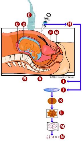
Like amniocentesis, chorionic villus sampling (CVS) is done to obtain a sample of the baby's tissue for determining genetic disorders. In contrast, CVS obtains tissue from the placenta (chorion) instead of fluid. Because the chorion is derived from the baby, not the mother, it bears the genetic characteristics of the baby. The procedure is similar to amniocentesis except that the tissue can be taken by inserting a needle through the abdomen or a sample tube through the cervix.

Like amniocentesis, CVS is done when the mother has one or more risk factors for genetic diseases. CVS can be done earlier than amniocentesis, usually in the latter part of the first trimester ( between weeks nine and 11). The results of CVS can be obtained sooner than amniocentesis because no culturing of the tissue is required. However, because CVS only gets tissue, some of the biochemical tests done with amniocentesis cannot be done with CVS. Also, CVS carries a higher risk (about 1 percent), mostly because it is a newer procedure relative to amniocentesis.

Advertisement

Fetal Monitoring

Fetal blood sampling can be done from 18 weeks to full term. In this procedure, fetal blood is obtained from the umbilical cord for analysis. Like amniocentesis, the doctor (aided by ultrasound) inserts a needle through the mother's abdomen into the umbilical cord and withdraws a blood sample. The blood sample is sent to a lab for analysis. The genetic results from fetal blood sampling can be obtained much faster than with amniocentesis because no tissue culturing is involved. In addition, this technique can be used to transfuse the fetus with compatible blood in the event that the baby's and mother's Rh factors do not match. The overall risk of the fetal blood sampling procedure is 0.5 - 1 percent.

Fetal monitoring is usually done in the third trimester (for high-risk pregnancies, pre-term labor or overdue babies) and during delivery. This test involves strapping an electronic monitor to the mother's abdomen that measures the electrical activities of the following:

Advertisement

  • fetal heart beat
  • mother's heart beat
  • mother's uterine muscle contractions

The baby's movements inside the uterus can be assessed and correlated with its heart rate. There are two types of test done:

  • Non-stress test - the baby's heart rate should increase when the baby moves (about 15 beats for 15 seconds at least twice in a 20 minute period).
  • Stress test - the baby's heart rate should increase when the uterus contracts. Uterine contractions are caused by infusing a medication (Pitocin) or by stimulating the mother's nipples.

These tests are used by obstetricians to determine how well the baby will handle the stress of delivery.

As you can see, there are a wide variety of ways to measure your baby's growth and development before it is born. Prenatal testing is completely normal during pregnancy and can often detect problems early, before they progress.

Advertisement

Advertisement

Loading...