How 3-D Bioprinting Works

Dr. Darryl D'Lima, an orthopedic specialist, works with a bioprinter he helped to develop located in the Shiley Center for Orthopedic Research & Education at Scripps Clinic. D' Lima has enlisted bioprinting in his cartilage regeneration research.
© Sandy Huffaker/Corbis

To make his eponymous monster, Victor Frankenstein needed body parts, but organ donation, as we know it, wouldn't emerge for another 135 years or so. And so the fictional doctor "dabbled among the unhallowed damps of the grave" and visited dissecting rooms and slaughterhouses, where he collected parts and pieces like some sort of ghoul.

Future Victor Frankensteins won't have to become grave robbers to obtain body parts. They won't even need bodies. Instead, we're betting they'll take advantage of a rapidly developing technology known as bioprinting. This offshoot of 3-D printing aims to allow scientists and medical researchers to build an organ, layer by layer, using scanners and printers traditionally reserved for auto design, model building and product prototyping.

Advertisement

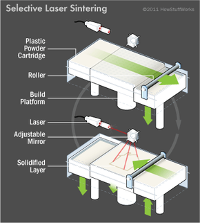
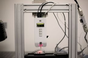
To make a toy using this technique, a manufacturer loads a substance, usually plastic, into a mini-fridge-sized machine. He also loads a 3-D design of the toy he wants to make. When he tells the machine to print, it heats up and, using the design as a set of instructions, extrudes a layer of melted plastic through a nozzle onto a platform. As the plastic cools, it begins to solidify, although by itself, it's nothing more than a single slice of the desired object. The platform then moves downward so a second layer can be deposited on the first. The printer repeats this process until it forms a solid object in the shape of the toy.

In industrial circles, this is known as additive manufacturing because the finished product is made by adding material to build up a three-dimensional shape. It differs from traditional manufacturing, which often involves subtracting a material, by way of machining, to achieve a certain shape. Additive manufacturers aren't limited to using plastic as their starting material. Some use powders, which are held together by glue or heated to fuse the powder together. Others prefer food materials, such as cheese or chocolate, to create edible sculptures. And still others -- modern versions of Victor Frankenstein -- are experimenting with biomaterials to print living tissue and, when layered properly in biotic environments, fully functioning organs.

That's right, the same technology that can produce Star Wars action figures also can produce human livers, kidneys, ears, blood vessels, skin and bones. But printing a 3-D version of R2-D2 isn't exactly the same as printing a heart that expands and contracts like real cardiac muscle. Cut through an action figure, and you'll find plastic through and through. Cut through a human heart, and you'll find a complex matrix of cells and tissues, all of which must be arranged properly for the organ to function. For this reason, bioprinting is developing more slowly than other additive manufacturing techniques, but it is advancing. Researchers have already built modified 3-D printers and are now perfecting the processes that will allow them to print tissues and organs for pharmaceutical testing and, ultimately, for transplantation.

Advertisement

The 3-D History of Bioprinting

Illustration of how one type of 3-D printing, selective laser sintering, works
HowStuffWorks.com

The promise of printing human organs began in 1983 when Charles Hull invented stereolithography. This special type of printing relied on a laser to solidify a polymer material extruded from a nozzle. The instructions for the design came from an engineer, who would define the 3-D shape of an object in computer-aided design (CAD) software and then send the file to the printer. Hull and his colleagues developed the file format, known as .stl, that carried information about the object's surface geometry, represented as a set of triangular faces.

At first, the materials used in stereolithography weren't sturdy enough to create long-lasting objects. As a result, engineers in the early days used the process strictly as a way to model an end product -- a car part, for example -- that would eventually be manufactured using traditional techniques. An entire industry, known as rapid prototyping, grew up around the technology, and in 1986, Hull founded 3D Systems to manufacture 3-D printers and the materials to go in them.

Advertisement

By the early 1990s, 3D Systems had begun to introduce the next generation of materials -- nanocomposites, blended plastics and powdered metals. These materials were more durable, which meant they could produce strong, sturdy objects that could function as finished products, not mere stepping-stones to finished products.

It didn't take long for medical researchers to notice. What's an organ but an object possessing a width, height and depth? Couldn't such a structure be mapped in three dimensions? And couldn't a 3-D printer receive such a map and then render the organ the same way it might render a hood ornament or piece of jewelry? Such a feat could be easily accomplished if the printer cartridges sprayed out biomaterials instead of plastics.

Scientists went on the hunt for such materials and by the late 1990s, they had devised viable techniques and processes to make organ-building a reality. In 1999, scientists at the Wake Forest Institute for Regenerative Medicine used a 3-D printer to build a synthetic scaffold of a human bladder. They then coated the scaffold with cells taken from their patients and successfully grew working organs. This set the stage for true bioprinting. In 2002, scientists printed a miniature functional kidney capable of filtering blood and producing urine in an animal model. And in 2010, Organovo -- a bioprinting company headquartered in San Diego -- printed the first blood vessel.

Today, the revolution continues. Taking center stage are the printers themselves, as well as the special blend of living inks they contain. We'll cover both next.

Advertisement

Just Like an Inkjet Printer, Sort Of

The idea of 3-D printing evolved directly from a technology everyone knows: the inkjet printer. Watch your HP or Epson machine churn out a printed page, and you'll notice that the print head, driven by a motor, moves in horizontal strips across a sheet of paper. As it moves, ink stored in a cartridge sprays through tiny nozzles and falls on the page in a series of fine drops. The drops build up to create an image, with higher-resolution settings depositing more ink than lower-resolution settings. To achieve full top-to-bottom coverage, the paper sheet, located beneath the print head, rolls up vertically.

The limitation of inkjet printers is that they only print in two dimensions -- along the x- and y-axes. A 3-D printer overcomes this by adding a mechanism to print along an additional axis, usually labeled the z-axis in mathematical applications. This mechanism is an elevator that moves a platform up and down. With such an arrangement, the ink head can lay down material from side to side, but it can also deposit layers vertically as the elevator draws the platform down and away from the print head. Fill the cartridge with plastic, and the printer will output a three-dimensional plastic widget. Fill it with cells, and it will output a mass of cells.

Advertisement

Conceptually, bioprinting is really that simple. In reality, it's a bit more challenging because an organ contains more than one type of material. And because the material is living tissue, it needs to receive nutrients and oxygen. To accommodate this, bioprinting companies have modified their 3-D printers to better serve the medical community.

Bioprinter Components

This bioprinter, located at the Shiley Center for Orthopedic Research & Education at Scripps Clinic in La Jolla, Calif., is displaying the temperature, pressure and drops/nozzle settings just above the three buttons. Could a future organ of yours be created on a bioprinter someday?
© Sandy Huffaker/Corbis

If you were to pull apart a bioprinter, as we'd love to do, you'd encounter these basic parts:

Print head mount -- On a bioprinter, the print heads are attached to a metal plate running along a horizontal track. The x-axis motor propels the metal plate (and the print heads) from side to side, allowing material to be deposited in either horizontal direction.

Advertisement

Elevator -- A metal track running vertically at the back of the machine, the elevator, driven by the z-axis motor, moves the print heads up and down. This makes it possible to stack successive layers of material, one on top of the next.

Platform -- A shelf at the bottom of the machine provides a platform for the organ to rest on during the production process. The platform may support a scaffold, a petri dish or a well plate, which could contain up to 24 small depressions to hold organ tissue samples for pharmaceutical testing. A third motor moves the platform front to back along the y-axis.

Reservoirs -- The reservoirs attach to the print heads and hold the biomaterial to be deposited during the printing process. These are equivalent to the cartridges in your inkjet printer.

Print heads/syringes -- A pump forces material from the reservoirs down through a small nozzle or syringe, which is positioned just above the platform. As the material is extruded, it forms a layer on the platform.

Triangulation sensor -- A small sensor tracks the tip of each print head as it moves along the x-, y- and z-axes. Software communicates with the machine so the precise location of the print heads is known throughout the process.

Microgel -- Unlike the ink you load into your printer at home, bioink is alive, so it needs food, water and oxygen to survive. This nurturing environment is provided by a microgel -- think gelatin enriched with vitamins, proteins and other life-sustaining compounds. Researchers either mix cells with the gel before printing or extrude the cells from one print head, microgel from the other. Either way, the gel helps the cells stay suspended and prevents them from settling and clumping.

Bioink -- Organs are made of tissues, and tissues are made of cells. To print an organ, a scientist must be able to deposit cells specific to the organ she hopes to build. For example, to create a liver, she would start with hepatocytes -- the essential cells of a liver -- as well as other supporting cells. These cells form a special material known as bioink, which is placed in the reservoir of the printer and then extruded through the print head. As the cells accumulate on the platform and become embedded in the microgel, they assume a three-dimensional shape that resembles a human organ.

Alternatively, the scientist could start with a bioink consisting of stem cells, which, after the printing process, have the potential to differentiate into the desired target cells. Either way, bioink is simply a medium, and a bioprinter is an output device. Up next, we'll review the steps required to print an organ designed specifically for a single patient.

Advertisement

Made-to-order Organs

The heart may be one of the easier organs to make with a bioprinter, said Stuart K. Williams, the head of the Cardiovascular Innovation Institute, in a 2013 interview with Wired.
Maciej Frolow/Photographer's Choice RF/Getty Images

When researchers built 3-D printers capable of depositing bioink and forming living masses of cells, they celebrated a major achievement. Then they immediately began to tackle the next big problem: How can bioprinting produce an organ for a specific person? To accomplish this, a medical team needs to collect data about the organ in question -- its size, shape and placement in the patient's body. Then team members need to concoct a bioink using cells taken from the patient. This ensures that the printed organ will be compatible genetically and won't be rejected once it's transplanted in the patient's body.

For simple organs, such as bladders, researchers don't print the living tissue directly. Instead, they print a 3-D scaffold made of biodegradable polymers or collagen. To determine the exact shape of the scaffold, they first build a 3-D model using computer-aided design (CAD) software. They usually define the exact x-, y- and z-coordinates of the model by taking scans of the patient using computerized tomography (CT) or magnetic resonance imaging (MRI) technology.

Advertisement

Next, researchers get the cells they need by taking a biopsy of the patient's bladder. They then place the cell samples in a culture, where they multiply into a population sufficiently large enough to cover the scaffold, which provides a temporary substrate for the cells to cling to as they organize and strengthen. Seeding the scaffold requires time-consuming and painstaking handwork with a pipette. It generally takes about eight weeks before such artificial bladders are ready for implantation. When doctors finally place the organ in the patient, the scaffold has either disappeared or disappears soon after the surgery.

The procedure above works because bladder tissue only contains two types of cells. Organs like kidneys and livers have a far more complex structure with a greater diversity of cell types. While it would be easy enough to print a scaffold, it would be almost impossible to recreate the three-dimensional structure of the tissue manually. A bioprinter, however, is ideally suited to complete such a time-consuming, detail-oriented task.

Advertisement

One, Two, Three, Print!

Here are the steps to print a complex organ:

  • First, doctors make CT or MRI scans of the desired organ.
  • Next, they load the images into a computer and build a corresponding 3-D blueprint of the structure using CAD software.
  • Combining this 3-D data with histological information collected from years of microscopic analysis of tissues, scientists build a slice-by-slice model of the patient's organ. Each slice accurately reflects how the unique cells and the surrounding cellular matrix fit together in three-dimensional space.
  • After that, it's a matter of hitting File > Print, which sends the modeling data to the bioprinter.
  • The printer outputs the organ one layer at a time, using bioink and gel to create the complex multicellular tissue and hold it in place.
  • Finally, scientists remove the organ from the printer and place it in an incubator, where the cells in the bioink enjoy some warm, quiet downtime to start living and working together. For example, liver cells need to form what biologists call "tight junctions," which describes how the cell membrane of one cell fuses to the cell membrane of the adjacent cell. The time in the incubator really pays off -- a few hours in the warmth turns the bioink into living tissue capable of carrying out liver functions and surviving in a lab for up to 40 days.

The final step of this process -- making printed organ cells behave like native cells -- has been challenging. Some scientists recommend that bioprinting be done with a patient's stem cells. After being deposited in their required three-dimensional space, they would then differentiate into mature cells, with all of the instructions about how to "behave." Then, of course, there's the issue of getting blood to all of the cells in a printed organ. Currently, bioprinting doesn't offer sufficient resolutions to create tiny, single-cell-thick capillaries. But scientists have printed larger blood vessels, and as the technology improves, the next step will be fully functional replacement organs, complete with the vascularization necessary to remain alive and healthy.

Advertisement

Uses for 3-D Organs

At the time of publication, surgeons hadn't implanted an organ printed from scratch into a human. That doesn't mean there haven't been successes. Replacing parts of the skeleton is one area being revolutionized by 3-D printing. Some dentists now take an intra-oral scan of a patient's teeth and send the scan to a lab that fashions a porcelain bridge using a 3-D printer. Prosthetic manufacturers also have changed their approach to designing artificial limbs. Now, many are able to print fairings -- prosthetic limb covers -- that mold perfectly to a person's anatomy, giving the wearer a more comfortable fit. These are just preludes to what the future may hold: printing entire bones for placement in the body. Doctors in the Netherlands have already created a lower mandible on a 3-D printer and implanted the jaw -- made from bioceramic-coated titanium -- in a patient suffering from a chronic bone infection.

Scientists have also successfully printed cartilaginous structures, such as ears and tracheas. To make the former, bioengineers take a 3-D scan of a patient's ear, design a mold using CAD software and then print it out. Then they inject the mold with cartilage cells and collagen. After spending some time in an incubator, the ear comes out, ready for attachment to the patient. A trachea can be made in a similar fashion. In 2012, doctors at the University of Michigan printed a sleeve, made from a 3-D model generated from a CT scan, to wrap and support a baby's trachea, which had been rendered weak and floppy by a rare defect.

Advertisement

The holy grail, of course, is a bioprinted organ, and skin -- the body's largest organ -- may be the first item on the list. Researchers at the Wake Forest Institute for Regenerative Medicine already have developed a complete system to print skin grafts. The system includes a scanner to map a patient's wound and a purpose-built inkjet printer that lays down the cells, proteins and enzymes necessary to form human skin. The goal is to build portable printers for use in field hospitals, where doctors can output skin directly onto patients.

Until these marvels come online, 3-D organs will play an important role in education and drug development. They might even factor into the development of food and clothing products (lab-grown meat and leather). Some medical schools have invested in 3-D printing technology to create surgical models of organs from CT or MRI images. This allows students to practice on hearts, livers and other structures that look and feel just like the real thing. Having access to such lifelike tissues also benefits pharmaceutical companies, which can test candidate drugs to see their effects. Organovo houses several printers capable of printing out three-dimensional models of liver, kidney and cancer tissues. These aren't full organs meant to live indefinitely. Instead, they're "organs on a chip" -- small, biologically active tissue samples designed to respond as native tissues would.

Perhaps one day, bioprinting will make anyone a Victor Frankenstein, capable of printing out organs, bones and muscles and assembling it all into a reasonable facsimile of a human. Then again, there's the issue of a nervous system. Even the best scanners, printers, inks and gels will fall short when it comes to recreating a thinking, dreaming brain. And without that, our efforts would leave us with a collection of anatomically correct, three-dimensionally accurate organs, but nothing to control them.

Advertisement

Lots More Information

Author's Note: How 3-D Bioprinting Works

I remember my first printer: a Brother typewriter hooked to a Commodore 64, followed by a daisy-wheel printer powered by an IBM PC. Hard to believe we might have bioprinters sitting on our desktops one day. If we do, I wonder where we'll go to get new bioink cartridges?

Related Articles

  • Atala, Anthony. "Printing a human kidney." TED Talks. March 2011. (Nov. 17, 2013)http://www.ted.com/talks/anthony_atala_printing_a_human_kidney.html
  • Banham, Russ. "Printing a Medical Revolution." T. Rowe Price. May 2012. (Nov 17, 2013)http://individual.troweprice.com/public/Retail/Planning-&-Research/Connections/3D-Printing/Printing-a-Medical-Revolution
  • Clark, Liat. "Bioengineer: the heart is one of the easiest organs to bioprint, we'll do it in a decade." Cardiovascular Innovation Institute. Nov. 21, 2013. (Nov. 25, 2013)http://cv2i.org/bioengineer-heart-one-easiest-organs-bioprint-well-decade/
  • Dutton, Gail. "3D Printing May Revolutionize Drug R&D." Genetic Engineering & Biotechnology News. Nov. 15, 2013. (Nov. 17, 2013)http://www.genengnews.com/gen-articles/3d-printing-may-revolutionize-drug-r-d/5062/
  • Fountain, Henry. "At the Printer, Living Tissue." The New York Times. Aug. 18, 2013. (Nov. 17, 2013) http://www.nytimes.com/2013/08/20/science/next-out-of-the-printer-living-tissue.html?
  • History.com Staff. "Organ Transplants: A Brief History." History.com. Feb. 21, 2012. (Nov. 17, 2013) http://www.history.com/news/organ-transplants-a-brief-history
  • Hsu, Jeremy. "3D Printed Organs May Mean End To Waiting Lists, Deadly Shortages." Huffington Post. Sept. 25, 2013. (Nov. 17, 2013)http://www.huffingtonpost.com/2013/09/25/3d-printed-organs_n_3983971.html
  • Hsu, Jeremy. "3D Printing: What a 3D Printer Is and How It Works." LiveScience. May 21, 2013. (Nov. 17, 2013) http://www.livescience.com/34551-3d-printing.html
  • Hsu, Jeremy. "Tiny 3D-Printed Organs Aim for 'Body on a Chip'." LiveScience. Sept. 16, 2013. (Nov. 17, 2013) http://www.livescience.com/39660-3d-printed-body-on-a-chip.html
  • Image Specialists. "How Inkjet Printers Work."http://www.image-specialists.com/ink_int_injet_printer.aspx
  • Leckart, Steven. "The Body Shop, Popular Science." Popular Science. August 2013.
  • Organovo. "The Bioprinting Process." (Nov. 17, 2013)http://www.organovo.com/science-technology/bioprinting-process
  • Pollack, Steven K. and Coburn, James. "FDA goes 3-D." FDAVoice. Aug. 15, 2013. (Dec. 13, 2013) http://blogs.fda.gov/fdavoice/index.php/2013/08/fda-goes-3-d/
  • Royte, Elizabeth. "What Lies Ahead for 3-D Printing?" Smithsonian Magazine. May 2013. (Nov. 17, 2013)http://www.smithsonianmag.com/science-nature/What-Lies-Ahead-for-3-D-Printing-204136931.html
  • Ungar, Laura. "Researchers closing in on printing 3-D hearts." USA Today. May 29, 2013. (Nov. 17, 2013) http://www.usatoday.com/story/tech/2013/05/29/health-3d-printing-organ-transplant/2370079/
  • Wake Forest Institute for Regenerative Medicine. "Using Ink-Jet Technology to Print Organs and Tissue." Aug. 2, 2013. (Nov. 17, 2013) http://www.wakehealth.edu/Research/WFIRM/Our-Story/Inside-the-Lab/Bioprinting.htm

Advertisement

Loading...