A Guide to Pregnancy Complications

MoThere are many possible pregnancy complications that prenatal care can help minimize. See more pregnancy pictures.
Publications International, Ltd.

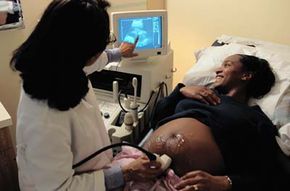
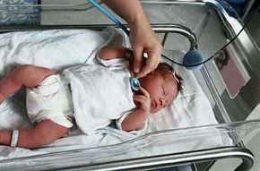
All parents hope and expect to experience a normal pregnancy and take home a healthy child. Although the majority of pregnancies and deliveries are uneventful, some involve complications that range from minor to life threatening -- for the mother, for the baby, or for both. Complications of pregnancy may develop gradually or suddenly and without warning. One objective of prenatal care is the prompt diagnosis and treatment of these complications before they worsen. In this article, we describe the most common complications of pregnancy, their causes and symptoms, and their treatment over the following sections:

  • Pregnancy Complications in Older Mothers In recent years there has been a trend toward having babies later in life. But more pronounced chronic diseases in the mother, among other conditions, can have an adverse affect on the developing fetus. Fortunately, prenatal care is similar for women of all ages, but this page will tell you some symptoms that women older than 35 should watch out for.
  • Pregnancy Complications in Teenage Mothers Most teenagers are not ready -- physically or emotionally -- to have children. As a result, birth weight is typically lower and babies of teens are more susceptible to certain illnesses. It will take an effort on the part of the teenage mom to ensure the health of her baby. This page tells you how to do it.
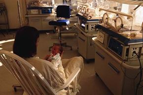
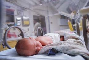
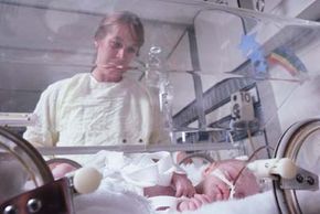
  • Preterm Birth About 11 to 12 percent of deliveries in the United States are classified as premature, which is a birth that occurs between the 20th and 36th weeks of pregnancy. This is a dangerous condition for the baby, which might not be able to survive outside of the womb. But modern medicine has helped many premature babies survive and grow up normally. On this page you'll learn why you must go to the hospital immediately if you suspect you're going into labor prematurely.
  • Postterm Birth If a baby still has not arrived two weeks past its due date, it may be in danger of malnutrition or even pneumonia. There is no danger to the mother, but a doctor may choose to induce labor for the baby's health. This page will tell you what happens in that case.
  • Ectopic Pregnancy An ectopic pregnancy is one in which the fertilized egg settles outside the uterus. This can be a very painful, and dangerous, condition for the mother, and if an ectopic pregnancy is detected the mother will be hospitalized immediately. This page will show you how to tell if you have an ectopic pregnancy, and what your doctor will do.
  • Stillbirths Stillbirth, the death of a baby before it is born, is becoming rare thanks to improved prenatal care. However, this tragic outcome of a pregnancy can happen, usually when the flow of nutrition from the placenta is cut off. This page will tell you how a doctor will detect a stillbirth.
  • Miscarriages About 15 percent of known births end in miscarriage, when a baby is born too early in the pregnancy to be viable. Although many doctors prescribe bed rest if they suspect a miscarriage is possible, most believe there is no way to prevent one. This page will tell you the different types of miscarriage and the factors that contribute to them.
  • Multiple Births Twins can double the joy of being a new mother, but they can also present health risks. The most common risk of a multiple birth is premature labor, and a breech birth -- with the baby coming out feet first rather than head first -- is common as well. Although the reasons for multiple births are not fully understood, this page will tell you what do to if you're carrying more than one fetus.
  • Placenta Complications A woman shouldn't experience any vaginal bleeding during pregnancy. If you do, it may be a sign of placenta previa or placental abruption. These are two conditions in which the placenta does not behave normally, and they almost always lead to a cesarean section. Find out more about these abnormalities on this page.
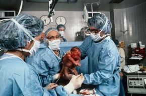
  • Cesarean Section About 30 percent of U.S. babies are born via cesarean section, in which a baby is removed through an incision in the mother's abdomen. There are many reasons why this procedure would be necessary, but it's always for the health of the baby or the mother and it's extremely safe. This page goes over the reasons and explains the two types of cesarean section.
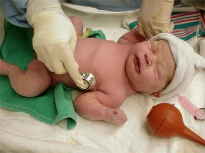
  • Birth Defects Birth defects can affect the head, face, eyes, mouth, hands, feet, and internal organs. Some minor birth defects can be corrected and leave no trace, but others are more severe and stay with the baby for life. This page details numerous birth defects, from cleft palate and clubfoot to spina bifida and Down syndrome.
  • Rh Incompatibility If a mother and fetus don't have compatible blood, there can be severe complications for the fetus. Furthermore, any fetus the mother carries in the future is at risk, too, unless a doctor takes the proper steps to sensitize the mother. Find out what Rh is, how it affects your pregnancy, and what your doctor will do about it on this page.
  • Deseases During Pregnancy The baby isn't the only one who's at risk. Pregnancy can cause health issues for the mother too, from simple swelling of the hands and feet to increased risk of heart and kidney disease. And existing conditions in the mother, such as diabetes, can endanger a fetus. Most mothers are healthy throughout their pregnancy, but this page will tell you how to stay that way.
  • Abnormalities of Labor Labor is a complicated process that can become difficult in several ways. If the baby and the umbilical cord are not positioned correctly, for example, the doctor may have to perform a cesarean section. Here you'll see what your doctor will be looking for and what he or she will do to correct anything that goes wrong.

This information is solely for informational purposes. IT IS NOT INTENDED TO PROVIDE MEDICAL ADVICE. Neither the Editors of Consumer Guide (R), Publications International, Ltd., the author nor publisher take responsibility for any possible consequences from any treatment, procedure, exercise, dietary modification, action or application of medication which results from reading or following the information contained in this information. The publication of this information does not constitute the practice of medicine, and this information does not replace the advice of your physician or other health care provider. Before undertaking any course of treatment, the reader must seek the advice of their physician or other health care provider.

Advertisement

Pregnancy Complications in Older Mothers

By medical tradition, an older mother is defined as a woman pregnant at the age of 35 or older. In recent years there has been a trend for women to delay childbearing until their 30s and to have additional children in their 30s and 40s. Although most older women experience successful pregnancies and deliver healthy babies, they may encounter a variety of problems.

Infertility

The inability to become pregnant -- called infertility -- is more common in older women. Because women begin to ovulate less frequently after age 30, the number of opportunities to achieve fertilization decreases as each year goes by. For example, the average woman at age 30 ovulates 13 times a year; by age 40, she may ovulate only 5 or 6 times a year.

Advertisement

Older women are also more likely to have problems with their reproductive organs, which may prevent pregnancy from occurring. Extensive endometriosis and uterine fibroids may make it impossible to become pregnant.

Chronic Illness

As we become older, we are more likely to develop chronic illnesses, for example, high blood pressure, diabetes, and glandular disorders, such as an under-active thyroid gland (hypothyroidism). Though these illnesses may not be serious or life-threatening to the nonpregnant woman, they may become more serious or even uncontrollable during pregnancy.

Furthermore, certain chronic illnesses of the mother are associated with an increased risk of miscarriage and stillbirth. Therefore, women with chronic illnesses need careful medical management during pregnancy.

Birth Defects

Older mothers are also at greater risk of having babies with severe birth defects caused by abnormalities of the baby's chromosomes.

Chromosomes are structures contained within all cells of the body, including the egg and the sperm. These chromosomes contain the genetic information passed on from parent to baby. Normally, the sperm and the egg each contain 23 chromosomes. When the sperm and the egg join, the resulting cell, which develops to form the baby, contains the normal chromosome number of 46.

In some cases, the egg may contain 24 chromosomes instead of the normal 23. If this egg combines with a normal sperm containing 23 chromosomes, the resulting fertilized egg contains 47 chromosomes -- an abnormal number. The baby that results from this fertilized egg then has 47 chromosomes in all the cells in its body.

In many cases, a woman carrying a fetus with an abnormal chromosome number miscarries, accounting somewhat for the higher miscarriage rate in older women. Those pregnancies that continue normally result in a fetus with any one of a number of physical abnormalities.

The most common condition associated with an abnormal chromosome number is Down syndrome, also called trisomy 21. Children with this condition are mentally retarded and may have serious abnormalities of the heart and digestive system. Children with Down syndrome have a characteristic facial appearance, with slanted eyes, heavy eyebrows, and a large, thick tongue.

Though Down syndrome babies may be born to mothers of any age, they are more frequently born to older mothers. At the age of 20, a mother has 1 chance in 1,667 of having a child with Down syndrome; at the age of 35, she has 1 chance in 385; at the age of 40, she has 1 chance in 106.

Treatment

The prenatal care of an older woman is generally like that of a woman in any other age group. If the woman has a history of high blood pressure, diabetes, or other chronic disorders, prenatal office visits may be more frequent than usual, especially during the last two months of pregnancy.

Mothers-to-be 35 years of age and older are offered prenatal testing to determine the presence of chromosome abnormalities in the fetus. Amniocentesis, the most commonly used test for detecting chromosome problems, is performed between the 16th and 18th weeks of pregnancy. Amniocentesis allows analysis of the fetal chromosomes to determine if there is an abnormal number. If an abnormality is discovered, the mother is given the option to terminate the pregnancy.

Labor and Delivery

Most older mothers experience normal labor and delivery. However, certain problems are more common in this age group, for example, placental abruption (premature separation of the placenta). Therefore, the rate of cesarean section is somewhat higher in older mothers.

There are also health risks for babies of mothers who become pregnant while very young. On the next page you'll learn about the complications of teenage pregnancy.

Pregnancy Complications in Teenage Mothers

There are special risks to a baby when the mother is not fully mature. Because most teenagers are not phsycially, emotionally, or financially ready to carry and care for a child, their babies tend to have low birth weight and are predisposed to a variety of illnesses. A teenage mother will need the full support of her family to live a healthy lifestyle for her and her baby.

Teen pregnancy has reached a 20-year low; however, teens give birth to approximately 500,000 babies each year. Teenagers often do not use contraceptives, and unfortunately, nearly two thirds of all teenage pregnancies are unintended.

Advertisement

The vast majority of teen mothers are not married, but few give up children for adoption or care by others. For this reason, the mothers often must drop out of school and cannot hold full-time employment. They must suddenly assume the responsibility of raising a child before they are ready, emotionally or financially.

Risks

Compared with mothers in older age groups, teenage mothers are at greater risk of having medical complications. Because the teenage mother is more likely to receive little or no prenatal care, she often becomes anemic and is more likely to develop preeclampsia, a severe condition associated with high blood pressure.

Vitamin deficiencies are more common, and the teenage mother's weight gain is likely to be inadequate. Since the teenage mother is still growing herself, she needs to eat properly not only for her own growth but for normal growth of the fetus.

Pelvic bones do not reach their maximum size until about the age of 18; therefore, the pelvis of the teenage mother may not have grown enough to allow vaginal delivery of a normal-size baby. For this reason, the incidence of cesarean section is higher in teenage mothers -- a baby that can be delivered vaginally when the mother is 20 is often too large to have been delivered vaginally when she was 14 years old.

Babies born to teenage mothers are more likely to die in the first year of life compared with babies born to mothers older than 20 years of age. Since the teenage mother is less likely to eat correctly during pregnancy, her baby often has a low birth weight (less than 51/2 pounds), making it more likely the baby will become ill.

Treatment

The teenage mother should be encouraged to seek prenatal care early in pregnancy, eat a nutritious diet, take prescribed vitamins and iron supplements, and engage in healthy physical activity. Though a supportive family can help the teenage mother cope with her new responsibilities, social service agencies may be needed to help her find ways to finish school and seek employment.

The Due Date

The average length of pregnancy is 40 weeks, or 280 days, from the first day of the last normal menstrual period. The due date, or expected date of delivery, for a pregnancy is calculated simply by adding nine months and seven days to the first day of a woman's last normal menstrual period. For example, if the first day of the last menstrual period was January 1, the expected date of delivery is nine months and seven days later -- on October 8. (Some physicians use the term expected date of confinement, or EDC, to describe the due date.)

In reality, the majority of women do not actually give birth on the due date. About 80 percent of babies are born within ten days of the due date-either ten days before or ten days after. As long as the delivery occurs between 37 and 42 weeks, the pregnancy is considered full term.

If the mother goes into labor before the due date, the baby could be in serious danger. In the next page, you will find out what to do if you go into premature labor.

Preterm Birth

If you go into labor before your 37th week, you should go to the hospital immediately. Find out why premature birth is dangerous to the baby.
Publications International, Ltd.

Premature birth can be very dangerous to a newborn because it might not be developed enough to survive outside of the womb -- for example, if the lungs are not yet able to breathe. There are several reasons it happens, most of which the mother has no control over. If you suspect that you are going into labor prematurely, you should go to the hospital immediately because labor can be stopped up to a certain point.

A preterm, or premature, delivery is defined as the birth of a baby between the 20th and 36th week of pregnancy. A baby born during this time is called premature. About 11 to 12 percent of deliveries in the United States are classified as premature; however, preterm deliveries are on the increase because more women are giving birth to multiple babies.

Advertisement

Causes

A preterm delivery occurs because the mother goes into labor too early. Though in most cases there is no clear reason, abnormally early labor is often associated with the following conditions:

  • Multiple pregnancy, such as twins or triplets
  • An abnormally shaped uterus that may crowd the fetus
  • Placenta previa
  • Placental abruption
  • Tobacco use
  • Untreated diseases of the thyroid gland in the mother
  • Other severe diseases in the mother, such as high blood pressure, diabetes, and kidney disease
  • High fever or severe infections in the mother

Contrary to popular belief, severe emotional trauma and physical injury, such as from a fall, are uncommon causes of premature labor. If a mother has premature labor in one pregnancy, she has a 25 percent chance of premature labor in the next pregnancy.

Complications

The major complication of preterm delivery is the birth of a baby who is unable to survive outside the mother's body. Though the baby's organs may all be correctly formed, his lungs may not be mature enough to allow him to breathe adequately after birth. Recent advances in the care of premature infants have allowed babies as small as 11/2 pounds to survive and grow up normally. Despite these advances, prematurity remains the leading cause of newborn death.

Treatment

If you suspect you are in labor before the 37th week of pregnancy, you should call your doctor and go to the hospital immediately. The doctor will check the cervix to determine if it has dilated. An electronic monitor may be used to detect contractions.

Several different drugs may be used to stop the uterine contractions of premature labor. These drugs are initially given intravenously or subcutaneously (under the skin), and some may be given orally once contractions have stopped. Once it has been determined that contractions have stopped, you may remain in the hospital for two to three days to make certain that contractions do not start again. Once you go home, a doctor will generally advise you to restrict physical activity, refrain from sexual intercourse, and rest in bed as much as possible.

Drugs used to stop preterm labor are more likely to be successful if the cervix has not dilated more than 4 centimeters (11/2 inches). If the cervix has dilated any more, most attempts to stop labor fail, and the baby will be delivered prematurely. This is one of the reasons why it is important to report to the hospital immediately if you suspect premature labor.

On the other side of the spectrum, a postterm birth can also put the newborn baby at risk. Find out how on the next page.

Postterm Birth

A postterm pregnancy occurs when the baby has not been delivered by the end of the 42nd week. There is no health risk to the mother, but after the 42nd week the fetus may be at risk of malnutrition. Also, if it passes fecal matter -- which usually does not happen until after birth -- and breathes it in, it may develop pnemonia. A doctor may choose to induce labor two weeks past the due date.

Cause

The cause of postterm pregnancy is not known. In most cases, however, it is believed that the mother misstated the exact date of her last menstrual period and the pregnancy was not postterm after all. If a woman has had one postterm pregnancy, she has a greater than average chance of this happening again in subsequent pregnancies.

Advertisement

Complications

Postterm pregnancy poses no health risk to the mother. However, as the placenta continues to age beyond the 42nd week of pregnancy, its ability to transmit oxygen and nutrients to the fetus may begin to decline. In some cases, this reduction in the transmission of oxygen and nutrients may be severe enough to cause the death of the fetus. If the fetus lives, at birth the baby commonly has a characteristic postterm appearance: wrinkled, cracking, peeling skin; long nails; abundant hair; and little fat tissue beneath the skin.

The postterm baby often passes fecal material called meconium into the amniotic fluid before delivery. If the baby sucks meconium into his lungs at the time of delivery, severe pneumonia may result.

Treatment

The usual treatment for postterm pregnancy is to periodically check the fetal heartbeat until labor starts on its own or is induced (started artificially). Labor is induced by administering the drug oxytocin (Pitocin) intravenously. Oxytocin stimulates uterine contractions similar to those of normal labor. A fetal monitor is generally used to detect any abnormalities of the fetal heartbeat. Most women with an induced labor experience a normal labor and delivery.

While postterm birth does not endanger the mother, ectopic pregnancy can be fatal. On the next page you'll learn what it is and how it affects the mother.

Ectopic Pregnancy

An ultrasound may reveal the location of an ectopic pregnancy.
Publications International, Ltd.

Ectopic (out of place) pregnancy occurs when the fertilized egg develops outside the uterus. The most common location of ectopic pregnancy is in one of the fallopian tubes (structures that extend about 41/2 inches from the ovaries to the uterus and through which the egg travels from the ovary to the uterus). An ectopic pregnancy that occurs in a fallopian tube is called a tubal pregnancy. On rare occasions, the pregnancy starts to develop in the ovary, on the cervix, or attached to the surface of a nearby organ.

An ectopic pregnancy is one in which the fertilized egg develops outside the uterus. One out of every 100 to 150 pregnancies is ectopic, most often in the fallopian tubes, but also in the ovary, on the cervix, or attached to another organ in the abdominal cavity.

Advertisement

Causes

The usual cause of ectopic pregnancy is an obstruction or narrowing of a fallopian tube that prevents the fertilized egg from passing through the tube into the uterus. This obstruction or narrowing is sometimes the result of inflammation and scarring from a previous pelvic infection caused by a Gonococcus or Chlamydia infection. Tubal infections caused by numerous other bacteria can also occur after miscarriage, after childbirth, or during the use of an IUD (intrauterine contraceptive device). If these infections are severe, blockage or narrowing of the tube may result.

Other less common causes of tubal obstruction or blockage include abdominal infections, such as appendicitis; pelvic tumors; and scar tissue formation after abdominal surgery.

Complications

An ectopic pregnancy may be fatal unless it is promptly treated. Left undetected it can rupture the tube enclosing it, leading to profuse bleeding into the abdomen. Ectopic pregnancies located in other areas, such as the ovary and cervix, can invade nearby blood vessels and cause massive bleeding. In earlier years, ectopic pregnancy was catastrophic, often leading to death. Today, with the advent of safe blood transfusions and better diagnostic methods allowing early diagnosis, death resulting from ectopic pregnancy is uncommon.

Symptoms

Symptoms of ectopic pregnancy usually appear two to four weeks after a woman has missed her menstrual period. Irregular spotting of blood from the vagina is one of the earliest symptoms. This is frequently followed by sharp, continuous pains on one side of the lower abdomen.

If the ectopic pregnancy ruptures, the woman usually experiences sudden, sharp, severe pain in the lower abdomen accompanied by rapid heartbeat and backache and, in some cases, fainting. However, the woman may experience no unusual symptoms even up to the point of rupture.

Diagnosis

During a pelvic examination, the doctor may discover a tender swelling on one side of the pelvis. Movement of the uterus or ovaries during this examination may cause pain. If there is a high suspicion of ectopic pregnancy, the woman is immediately hospitalized.

When an ectopic pregnancy is a possibility, an ultrasound of the pelvic structures may reveal the location of the ectopic pregnancy. Blood that has leaked from the ectopic pregnancy into the abdominal cavity may be detected by inserting a hollow needle through the wall of the vagina beneath the cervix and drawing off blood or by an examination using a pelvic ultrasound. The diagnosis is confirmed by inserting a laparoscope (a lighted, tube-like instrument) into the abdominal cavity through a small incision made below the navel. This allows the doctor to look directly at the pelvic organs and precisely locate the ectopic pregnancy.

Treatment

Treatment for ectopic pregnancy depends on several factors, including the size of the embryo and whether it has caused a rupture or not. If the embryo is small and the woman is not experiencing significant pain or bleeding, ectopic pregnancy may be treated using methotrexate, a drug that interferes with the growth of the embryo and causes it to be reabsorbed into the body.

Methotrexate is administered as an injection, and the woman must be closely monitored for complications and success of the treatment. If the treatment fails, the embryo is large, or the woman is experiencing significant pain or bleeding, surgical removal is necessary.

A woman who has had one ectopic pregnancy has a 15 percent chance of having a second one. This does not mean she should not try to become pregnant again, but when she does try, she should be especially watchful for symptoms of ectopic pregnancy. She should see her doctor as soon as she suspects she is pregnant so the location of the embryo may be determined.

Even if the embryo develops normally, the pregnancy can still end in tragedy. On the next page you'll learn about the factors that lead to a stillbirth.

Stillbirths

Stillbirths, which come in the second or third trimester, are thankfully growing less common, but it is always sad news for the family when it happens. However, the conditions that can lead to a stillbirth, such as placenta complications and twisting of the umbilical cord, are often things that the mother has no control over. And the silver lining is that a stillbirth typically does not endanger the life of the mother.

The death of the fetus at some time between the 20th week of pregnancy and birth is called stillbirth -- in medical terms, an intrauterine fetal demise. This tragic outcome of pregnancy is uncommon today because of better prenatal care and improved methods of diagnosing and treating abnormal pregnancies.

Advertisement

Causes

The primary cause of stillbirth is interruption of the normal flow of oxygen and nutrients from the mother to the fetus via the placenta and the umbilical cord. Conditions that may adversely affect the placenta and cause stillbirth include toxemia, chronic high blood pressure, diabetes, placenta previa, and placental abruption. Less commonly, a problem with the umbilical cord, such as twisting or breakage of a blood vessel, may cut off the flow of blood to the fetus and lead to stillbirth.

Certain abnormalities of the fetus -- including erythroblastosis; severe abnormalities of the heart, kidneys, and nervous system; and even fetal heart attack -- may lead to stillbirth. It is extremely rare for an injury to the mother to cause stillbirth.

Complications

The death of the fetus within the uterus usually does not jeopardize the mother's health. The body generally has no reaction to fetal death except for loss of weight. Uncommonly, death of the fetus may cause abnormalities of the mother's blood clotting system, but only after the fetus has been dead for several weeks.

Diagnosis

Fetal death is usually brought to the doctor's attention by the woman's reporting that she has not felt the fetus move for a day or two. This absence of fetal movement is significant only in the last few months of pregnancy; before this, failure to note fetal movement for a day or two is normal. If the doctor fails to detect a heartbeat, confirmation of the diagnosis of stillbirth is sought with either an electronic heartbeat monitor or ultrasound.

Treatment

Spontaneous labor may begin any time from a few hours to up to 60 days after the death of the fetus. When labor does occur, it is usually normal. Today, most doctors choose to induce labor and deliver the fetus as soon as possible after the diagnosis of fetal death. This is accomplished either by administering the drug oxytocin intravenously or giving drugs called prostaglandins as vaginal suppositories.

A pregnancy can also end tragically in miscarriage, and about 15 percent of births do. On the next page you'll learn what causes miscarriage and what the different types are.

Miscarriages

A miscarriage (in medical terms, a spontaneous abortion) is the expulsion from the uterus of the fetus and placenta before the beginning of the 20th week of pregnancy. At that point, the fetus is not developed enough to survive outside the uterus on its own. (After the 20th week of pregnancy and before the 36th week, expulsion of the fetus and placenta is considered premature delivery.) Most miscarriages occur within the first 14 weeks of pregnancy.

It is impossible to know how many miscarriages occur during the first month of pregnancy, before many women may even realize they are pregnant. The only indication may be a slightly late menstrual period with a heavier than normal flow. However, about 15 percent of known pregnancies end in miscarriage.

Advertisement

Types

There are different categories of miscarriages.

  • One of every five pregnant women experiences a threatened miscarriage when she bleeds vaginally during the first three months. Although it may indicate that a spontaneous abortion will eventually occur, it is often no more than a threat, and the pregnancy continues normally.
  • An inevitable miscarriage occurs when the woman begins to bleed and the cervix dilates; it is then only a matter of time before the contents of the uterus are expelled.
  • A missed miscarriage occurs when the fetus dies and the placenta stops growing; this may occur several days to weeks before the contents of the uterus are naturally expelled.
  • An incomplete miscarriage occurs when only part of the uterine contents has been expelled.
  • A complete miscarriage occurs when all of the uterine contents have been naturally expelled.

Causes

The reason a miscarriage occurs is not always known, but in many cases it is believed that a fetus aborts because it is not developing normally. Several factors can contribute to abnormal fetal development, including the following:

  • Abnormalities in the father's sperm
  • Abnormalities in the egg
  • Disease in the mother, most notably rubella (German measles), severe heart or kidney disease, diabetes, or thyroid disease
  • Abnormalities in the uterus
  • The mother's use of certain drugs
  • The mother's exposure to toxic substances or certain environmental pollutants

Contrary to popular belief, severe emotional trauma or stress, automobile accidents, and simply falling rarely, if ever, cause miscarriage.

The expulsion of the fetus because of an abnormality is thought to be a chance event, usually not due to a defect in either parent. Of women who miscarry once, most (80 percent) have a successful subsequent pregnancy.

Although it is uncommon, some women miscarry three or more times in a row; they are called habitual aborters. When this occurs, the physician conducts a thorough evaluation of both the woman and her partner to determine the cause, if any. Frequently, a chromosome abnormality in one parent or an abnormality of the uterus is found. If an abnormality of the uterus is the cause of miscarriage, corrective surgery may be done, and a successful pregnancy often results. If the cause of miscarriage is a chromosome abnormality in either parent, the problem cannot be corrected.

Complications

Miscarriage is rarely life-threatening for the mother, especially if diagnosed and treated promptly. Though blood loss may occur, it is generally not enough to cause serious problems.

Symptoms

The symptoms of miscarriage are vaginal bleeding (from a few drops to a heavy flow) and uterine cramps (either dull and constant or sharp and intermittent) felt in the lower part of the abdomen or back. The bleeding can start suddenly or follow a brownish discharge. A solid clot of blood or tissue may pass from the vagina. If possible, this should be saved for the doctor, who may be able to examine it and determine the cause of the bleeding.

A pregnant woman who starts bleeding or experiences abdominal pain should contact her doctor immediately.

Treatment

If a threatened miscarriage is diagnosed, the doctor generally directs the woman to rest in bed, avoid heavy lifting and pushing, and abstain from sexual intercourse. Though it is traditional for doctors to give this advice, many physicians feel little can be done to stop or avert a miscarriage.

After an inevitable, incomplete, or missed miscarriage, any tissue remaining in the uterus causes continued bleeding and possibly infection. To remove any retained tissue, the doctor performs a D & C (dilatation and curettage). This is a surgical procedure in which the cervix is dilated by means of tapered metal dilators, and the contents of the uterus are scraped out with suction and a sharp instrument called a curette.

If you are carrying more than one fetus, you also might not carry to full term. On the next page find out why multiple births increase the liklihood of premature labor.

Multiple Births

Multiple births can be a joyous occasion, but they are not without peril.
Publications International, Ltd.

Twins and triplets are often seen as an additional blessing on a growing family. But multiple births also can cause complications, the most serious of which is premature birth. Because the babies may not be ready to live outside the mother's womb, treatment of multiple births mostly involves preventing premature labor, and lots of bed rest is a common method.

The number of multiple births is increasing as women wait until they are older to have a baby and use fertility drugs to conceive. According to the National Center for Health Statistics, between 1980 and 2000, the number of twin births increased 74 percent, and the number of triplets, quadruplets, and other higher order births increased fivefold.

Advertisement

Multiple births occur more commonly in older women; women who have had at least one previous pregnancy; women who have a family history of multiple pregnancy on the mother's side of the family; and women who have taken fertility drugs to stimulate ovulation (the expulsion of an egg from the ovary).

Types

There are two types of twins. One type starts from a single egg, which divides in two very early after fertilization. Because a single egg is fertilized by a single sperm before this division, the two offspring will be of the same sex and alike in skin, hair, and eye color and in general appearance. They are identical twins.

An extremely rare form of identical twinning is called conjoined, or Siamese, twinning. In these unfortunate cases, the twins are attached to each other at the head, chest, abdomen, or back. Surgical separation may be successful but often results in the death of one or both babies.

The other type of twinning is the result of fertilization of two eggs by two different sperm. Twins of this type -- called fraternal twins -- may be of the same sex or of opposite sexes and bear no greater resemblance to each other than any other brothers and sisters. Most twins that result from the use of fertility drugs are of this type.

Approximately one third of twins are identical, and two thirds are fraternal.

One or more eggs may be involved in other forms of multiple pregnancy. For example, quadruplets may result from one, two, three, or four eggs.

Causes

In most cases of multiple birth, the cause is unknown. Why certain families have many sets of twins is also not understood. Fertility drugs used to stimulate ovulation are often associated with multiple births -- sometimes up to eight fetuses.

Diagnosis

Doctor and patient alike often suspect multiple pregnancy when the woman's uterus grows more rapidly than one would expect with one baby. Confirmation comes when the doctor hears two separate fetal heartbeats or an ultrasound shows two or more fetuses.

Complications

The major complication of multiple pregnancy is premature labor, with the delivery of small, premature babies who are not yet ready to live outside the mother's body. In general, the more fetuses a woman is carrying, the earlier in pregnancy she is likely to go into labor. Other complications that may affect the mother are anemia and preeclampsia. Women with multiple pregnancies are very carefully monitored.

Treatment During Pregnancy

The doctor's special care of a woman with a multiple pregnancy aims at preventing premature labor and detecting and treating anemia and preeclampsia. When multiple pregnancy is diagnosed, the woman is advised to avoid strenuous activity and rest in bed as much as possible. She may even be admitted to the hospital weeks before the expected delivery date. Prenatal visits are more frequent than usual (at least once a week) so the doctor can detect the early stages of premature labor and preeclampsia.

Labor and Delivery

The position of the twins in the uterus is quite variable: both babies may be head first, the first baby may be head first and the second may be breech (buttocks or legs first), the first may be breech and the second may be head first, or both may be breech. If both are head first, doctors usually allow a vaginal delivery. When the babies are in other positions, however, a cesarean section is usually performed to ensure a safe delivery of both babies. Because of the serious problems that may be encountered in the vaginal delivery of other types of multiple pregnancies, most pregnancies involving three or more babies are delivered by means of cesarean section.

A cesarean section may be necessary for multiple births, and is almost always necessary when there are complications with the placenta. Read on to learn about these conditions.

Placenta Complications

Vaginal bleeding during pregnancy can be a sign of a problem with the placenta. Find out what to do about placenta previa and placental abruption.
Publications International, Ltd.

From the time of the last normal menstrual period to the time of labor, a pregnant woman should not experience any bleeding from the vagina. During the first half of pregnancy, the most serious causes of bleeding are miscarriage and ectopic pregnancy; during the last half of pregnancy, the most serious causes are placenta previa and placental abruption.

Placenta Previa

In placenta previa, the placenta is located low in the uterine cavity, partially or completely covering the opening of the cervix. As the lower portion of the uterus stretches and dilates during the latter weeks of pregnancy, portions of the placenta may be torn from their attachment to the wall of the uterus. This leads to variable amounts of bleeding, ranging from light to profuse.

Advertisement

Causes

The exact cause of placenta previa is unknown. It is the leading cause of vaginal bleeding during the second and third trimesters.

Symptons

The major symptom of placenta previa is painless vaginal bleeding. This bleeding may begin as early as the 24th to 26th week of pregnancy, though it is more common during the last 4 or 5 weeks. The blood is usually bright red, indicating the bleeding is fresh. This bleeding occurs without any previous injury (for example, from a fall). In some cases, the blood flow is light (referred to as spotting). Abdominal pain or cramping generally does not accompany the bleeding unless the woman is in labor.

Diagnosis

Any vaginal bleeding during pregnancy is abnormal and should be reported to the doctor immediately. The doctor orders an ultrasound image of the mother's abdomen to locate the placenta. In most cases of placenta previa, the ultrasound shows the placenta covering part or all of the opening of the cervix.

Complications

Bleeding from a placenta previa may be extremely heavy, leading to severe blood loss from the mother. This bleeding stops only after the baby has been delivered and the placenta has been removed. Unless treatment is rapid in the case of heavy bleeding, death of both the mother and the baby may result.

Treatment

Since the placenta covers part or all of the opening of the cervix, a normal vaginal delivery cannot be attempted since extreme hemorrhage will occur. For this reason, babies of most mothers with placenta previa are delivered by means of cesarean section.

Once placenta previa has been diagnosed, the doctor recommends the best treatment -- either delayed or active. Delayed treatment involves admitting the woman to the hospital, keeping her on bed rest, and closely monitoring her for any recurrence of bleeding. The purpose of delayed treatment is to give the fetus time to mature so that it can survive outside the mother's body. Once the maturity of the fetus has been established, delivery is by means of cesarean section. In the case of active treatment, the baby is delivered by means of cesarean section as soon as possible.

The degree of vaginal bleeding usually dictates the type of treatment. If vaginal bleeding is slight and the fetus is not yet mature, the doctor generally recommends delayed treatment. If vaginal bleeding is heavy, a cesarean section is performed immediately to save the life of the mother and the baby, even if the baby is premature.

Placental Abruption

After delivery of a baby, the uterus begins to contract, separating the placenta from its wall and pushing it into the vagina. In the case of placental abruption, the placenta begins to separate from the uterine wall before the delivery of the baby. This occurs to some degree in about 1 of every 150 pregnancies.

Causes

The exact cause of placental abruption is unknown. However, a number of conditions are commonly associated with placental abruption, including high blood pressure in the mother, severe trauma to the mother's abdomen, cigarette smoking or drug use, and advanced age of the mother.

Symptoms

The major symptoms of placental abruption are vaginal bleeding and strong, continuous pain over the uterus and sometimes across the back. Bleeding may be as slight as a tablespoon or extremely heavy. In some cases, no bleeding is visible, and the only symptom is pain. If blood loss is great, the woman experiences rapid heartbeat, difficulty breathing, fainting, and even shock.

Diagnosis

The diagnosis of placental abruption is made on the basis of the woman's history and the doctor's findings of blood in the vagina and a tender, painful uterus. No specific tests can diagnose placental abruption. In most cases, even the findings on ultrasound are normal.

Complications

Bleeding associated with a placental abruption may be extremely heavy and, without rapid treatment, may lead to death of both the mother and the fetus. Even if the mother does not bleed heavily, the placenta may separate from the wall of the uterus to an extent that the fetus does not receive a sufficient oxygen supply to survive. In some cases, the mother's blood may fail to clot normally, leading to even more profuse vaginal bleeding.

Treatment

The treatment of placental abruption may be either delayed or active. If vaginal bleeding is slight or has stopped and the fetus remains unharmed, the mother is admitted to the hospital for observation until all signs of placental abruption have stopped. If, on the other hand, vaginal bleeding is heavy or the fetus shows signs of lack of oxygen, delivery must occur quickly. A mother who is already in labor is monitored closely and may expect to give birth vaginally if she and the fetus remain stable. In the case of heavy bleeding, a cesarean section is performed immediately. Before or after delivery, the mother may require a blood transfusion if she experiences severe blood loss.

Each of these conditions can lead to a cesarean section. Continue reading to find out more about this extremely safe procedure.

Cesarean Section

Cesarean section is necessary when a normal birth would endanger either the mother or the baby. This can be due to the size and position of the baby, the presence of bacteria, or because of multiple births. There are two types of cesaren section, but modern medicine has made both of them extremely safe procedures.

Cesarean section is the delivery of a baby by cutting through the abdominal wall and uterus and removing the baby through these incisions. Almost 30 percent of all births in the United States are by cesarean section.

Advertisement

Reasons

Cesarean section is performed when delivery of the baby is necessary and when a vaginal delivery would cause injury to either the mother or the baby. Some of the specific reasons for a cesarean section include the following:

  • To save the baby's life when problems with the placenta or the umbilical cord cut off the blood supply to the baby (This event is often detected when abnormal heart rate patterns appear on a fetal monitor.)
  • To deliver the baby if the mother is unable to give birth after a long labor, in most cases because the baby is too large to pass through the pelvis
  • To prevent infection of the baby from dangerous bacteria in the surrounding amniotic fluid or a dangerous vaginal or cervical infection
  • To prevent injury to the baby during a breech birth, when the baby would emerge buttocks or feet first through the vagina rather than head first (Many obstetricians believe that all breech births in first-time mothers and all premature breech births require cesarean delivery.)
  • To treat disease of the mother or the baby that can be treated better if birth occurs as soon as possible
  • To prevent the chance of rupture of the uterus during labor if the mother has had a previous cesarean section, especially of the classical type
  • To deliver multiple pregnancies involving three or more fetuses

Types

Two types of cesarean section are performed:

  • In the classical cesarean section, a vertical cut is made directly down the center of the uterus in its thick upper section. The incision on the skin may be horizontal or vertical. This operation is generally performed only if the baby is lying in an abnormal position or if the placenta is in an abnormally low position in the cavity of the uterus. With this type of cesarean section, more bleeding occurs than with other methods, the incision is more difficult to repair, and the uterus is more likely to rupture during a future pregnancy. For these reasons, the classical cesarean section is seldom used today unless there are specific reasons for its use.
  • The lower-segment cesarean section is the more commonly performed operation. Here, a horizontal incision is made in the lower, thinner portion of the uterus. The incision made on the skin may be smile-shaped (the "bikini" incision) near the lower part of the abdomen.

Risks

To minimize risks associated with a cesarean section, the operation should be performed only by a skilled obstetrician, accompanied by an anesthesiologist to administer anesthesia to the mother. In some cases, a pediatrician may also participate to take care of the baby after birth.

Risks associated with cesarean section include possible infection, excessive bleeding, and dangerous blood clots that may enter the blood circulation.

Further, the operation may be inconvenient for the mother, requiring her to stay in the hospital at least two or three days instead of going home with the baby soon after a vaginal delivery. And although the use of general anesthesia is now uncommon with cesarean section, if it was used, a woman may not be able to see the baby for a number of hours after delivery. (Breast-feeding following cesarean section should not present a problem if adequate pain relief is provided and maintained for the mother.)

She will have pain from the operative incision, and once she arrives home, she will need to restrict her activity for a while. It takes up to six weeks for complete healing of the incision.

Of course, all these inconveniences are small if the result is a healthy mother and a healthy baby. And today, fortunately, cesarean section is an extremely safe operation because of improved antibiotics and better anesthesia.

Effects on Parents

In the past, women undergoing cesarean section have been separated from their husbands during surgery, and as a result have often expressed negative feelings of isolation and inadequacy for having failed to accomplish a vaginal delivery. To assist these parents and prevent this from happening to others, many childbirth instructors provide couples with more information about cesarean section.

In many hospitals, husbands are allowed to remain with their wives during the operation. Generally, allowing fathers in the operating room has not caused problems, and women who have undergone cesarean section with their husbands at their sides have shared and enjoyed the birth experience in a far more positive way.

Vaginal Birth After Cesarean Section

In the past, a firm rule in obstetrics was "once a section, always a section." More recently, obstetricians have found that many women with a previous cesarean section can have a safe vaginal delivery in a subsequent pregnancy. To minimize the risks of rupture of the uterine scar during labor, certain criteria must be met: The previous cesarean must have been only the lower-segment type; the mother's pelvis must be of normal size; and the indication for the previous cesarean should not exist with the present pregnancy.

While cesarean sections do not typically affect a baby long-term, birth defects can be lifelong conditions. On the next page you'll learn what some of the more common birth defects are.

Birth Defects

All expectant parents dream of having normal, healthy children. Unfortunately, sometimes this does not happen. Although most babies are born normal and whole, about 3 percent are born with some form of abnormality. Fortunately, about half of these imperfect babies have only minor defects that can be easily corrected, leaving no trace. In the other half, however, the defect may be severe and even life-threatening.

Types

Birth defects, also called congenital anomalies, can affect nearly every organ of the baby's body. In some cases, these defects are visible on the surface of the body; in other cases, the defects involve internal organs, such as the heart or intestines. Another type of defect, called an inborn error of metabolism, is not visible but rather is an abnormality of the chemical system of the body in which normal chemical reactions in certain organs cannot occur.

Advertisement

The following is a list of the most common serious birth defects:

  • Head and face: Hydrocephalus (an abnormally large head due to accumulation of fluid), microcephaly (an abnormally small head), cleft lip and cleft palate (an abnormal opening in the lip and the roof of the mouth)
  • Heart: Congenital heart disease (a variety of different defects in the valves and walls of the heart)
  • Lungs: Absence of one lung, abnormal lung development
  • Stomach and intestines: Abnormal blockage
  • Abdominal wall: An opening through the navel or a large defect that causes the intestines to protrude
  • Kidneys: Absence of one or both kidneys, large cysts in the kidneys, developmental abnormalities of the kidneys (such as variations in structure)
  • Back: Spina bifida (an abnormal opening in the backbone in which nerves are contained in a thin, saclike structure)
  • Arms and legs: Absence of fingers or toes, fused fingers or toes, total absence of an arm or leg, clubfoot (an abnormal inward turning of the foot)

Causes

Little is known about the cause of most congenital anomalies. Heredity plays a role in some cases; some families have several members with similar abnormalities. Certain drugs, environmental pollutants, toxins, and high doses of radiation cause specific abnormalities. Certain infections in the mother -- the most significant of these is German measles (rubella) -- may cause multiple severe abnormalities. The mother's heavy drinking during pregnancy can cause fetal alcohol syndrome. In most cases, however, the exact reason for the abnormality is not known and probably derives from a disturbance in the normal organ development of the fetus.

Diagnosis

The ability to detect congenital abnormalities before birth is a relatively recent medical achievement. In addition to allowing some couples to terminate an abnormal pregnancy, it has enabled others who may be at risk of having abnormal offspring to continue a pregnancy knowing the fetus is normal.

During the first prenatal visit, the doctor obtains a thorough history of both parents, including age, race, ethnic background, and previous abnormal conditions in the family. This information is used to determine if testing for an abnormal fetus is necessary. The following tests detect specific abnormalities:

  • Ultrasonography: detects hydrocephalus, microcephaly, spina bifida, tumors, heart defects, intestinal blockage, and absence of an arm or leg.
  • Blood tests: The alpha-fetoprotein (AFP) test (the most commonly used blood test to detect fetal abnormality) helps detect spina bifida.
  • Amniocentesis: detects chromosome abnormalities, spina bifida, and nearly 100 chemical disorders in the fetus.
  • Chorionic villus sampling (CVS): detects the same chromosomal abnormalities as amniocentesis, but the test can be performed earlier in the pregnancy.

Treatment

Once an abnormality of the fetus has been detected, the parents must make a decision to either terminate or continue the pregnancy. However, the many remarkable advances in medical and surgical care can wholly cure many birth abnormalities considered very serious a few decades ago. Many heart defects, intestinal obstructions, and abnormalities of the abdominal wall can be corrected surgically, and those children born with them can expect to lead normal and healthy lives.

A pediatrician and a surgeon should describe to the parents the extent of the fetal abnormality and the chances of correcting it. Research currently underway may eventually enable surgical correction of certain types of congenital abnormalities while the fetus is still in the uterus.

Future Pregnancies

Fortunately, in most cases, birth defects do not repeat themselves in future pregnancies. Parents with one abnormal child have an average risk of less than 5 percent of having another child with the same abnormality. If, however, the defect is thought to be inherited from one parent, a specific pattern of this abnormality may be repeating itself. Before the parents of an abnormal child plan another pregnancy, they should meet with a doctor skilled in genetics and fetal abnormalities to discuss the risks of abnormalities appearing again.

Effect on the Parents

The birth of a physically abnormal child can produce feelings of shock, disbelief, sadness, and even anger in the parents. All members of the family are in some way affected by the birth. The parents must create a mutual support system to cope with this crisis. Gradually, the parents resolve their grief and become as closely attached to this child as they would to any other.

Genes, disease, and alcohol use can all contribute to abnormal development of a fetus. Continue reading to find out how a fetus also can be affected if its blood is incompatible with the mother's.

Rh Incompatibility

Because not everyone has the substance know as the Rh factor, it becomes an issue during pregnancy. While it may seem counterintuitive, the problem arises when the mother is Rh-negative and her fetus is Rh-positive, and the problem can affect a future fetus as well. A simple injection can guard against life-threatening conditions in a fetus.

Approximately 90 percent of people are born with a substance on their blood cells known as the Rh factor, so called because a similar substance is found on the blood cells of rhesus monkeys. Individuals who have this factor on their blood cells are called Rh-positive; those without it are called Rh-negative. The presence of the Rh factor is inherited from one's parents.

Advertisement

Problems can occur in the fetus if the mother is Rh-negative and the fetus is Rh-positive. Normally, some fetal blood escapes from the placenta and enters the mother's blood. If both the mother and fetus are Rh-positive, this leakage of fetal blood causes no problems. However, since this fetal blood containing the Rh factor is a foreign substance to a mother who is Rh-negative, the mother's body forms antibodies against the Rh factor.

The body normally forms antibodies in response to foreign substances -- usually bacteria and viruses that may be harmful. These antibodies act by destroying the foreign substance, thus protecting the body against their harmful effects. A person who forms antibodies against a substance is called sensitized to that particular substance.

If the mother forms these antibodies against Rh-positive blood, the antibodies cross from the mother into the fetus and start destroying fetal blood cells. This leads to a serious condition in the fetus called erythroblastosis, which may lead to anemia, heart failure, and even stillbirth.

When an Rh-negative mother becomes sensitized, it is usually at the time of the birth of her first Rh-positive baby. This baby is generally unaffected. But the mother keeps her antibodies against Rh-positive blood for life. If she has another pregnancy with an Rh-positive fetus, this fetus may be affected.

During early pregnancy, the doctor performs a blood test to determine if the mother has Rh-positive or Rh-negative blood. If the mother is Rh-negative, the father is also tested to determine his Rh factor. If he is Rh-positive, the baby may be Rh-positive as well.

As a precaution, to prevent the mother from becoming sensitized to Rh-positive blood, her doctor may recommend she receive an injection of a substance called human anti-Rh immune globulin at 28 weeks of pregnancy. If her baby is found at birth to be Rh-positive, the mother will receive another injection within two days of delivery.

The human anti-Rh immune globulin prevents the mother's body from developing Rh antibodies that would cause complications. An injection may also be given if the mother has an amniocentesis, a miscarriage, or an ectopic pregnancy. If it is found that the mother has already developed Rh antibodies, the pregnancy is closely monitored to determine if blood transfusions are necessary.

The Rh factor is an issue only during pregnancy. On the next page you'll learn about diseases that are either exclusive to pregnancy or more pronounced during this time.

Toxemia of Pregnancy

Several diseases are more pronounced during pregnancy, and several more affect only pregnant women. Diabetes is no longer a life-threatening complication, but can lead to abnormally small babies.
Publications International, Ltd.

The majority of women enter pregnancy healthy and remain so until delivery of the baby. Aside from a variety of minor problems -- for example, backache, morning sickness, and constipation -- the woman generally tolerates the many changes in her body that result from pregnancy. In some cases, however, either the mother enters pregnancy with a medical problem or a problem develops during pregnancy. Some of these conditions affect only the mother, while others may affect both mother and baby.

Toxemia of Pregnancy

Toxemia of pregnancy is a severe condition that sometimes occurs in the latter weeks of pregnancy. It is characterized by high blood pressure; swelling of the hands, feet, and face; and an excessive amount of protein in the urine. If the condition is allowed to worsen, the mother may experience convulsions and coma, and the baby may be stillborn.

Advertisement

The term toxemia is actually a misnomer from the days when it was thought that the condition was caused by toxic (poisonous) substances in the blood. The illness is more accurately called preeclampsia before the convulsive stage and eclampsia afterward.

Causes

The causes of preeclampsia and eclampsia are not clearly understood. They tend to develop more often in women who are having their first baby, especially teenage mothers and women from lower socioeconomic groups. One theory proposes that certain dietary deficiencies may be the cause of some cases. Also, there is the possibility that some forms of preeclampsia and eclampsia are the result of deficiency of blood flow in the uterus.

Symptoms

The symptoms of toxemia of pregnancy (which may lead to death if not treated) are divided into three stages, each progressively more serious:

  1. Mild preeclampsia symptoms include edema (puffiness under the skin due to fluid accumulation in the body tissues, often noted around the ankles), mild elevation of blood pressure, and the presence of small amounts of protein in the urine.
  2. Severe preeclampsia symptoms include extreme edema, extreme elevation of blood pressure, the presence of large amounts of protein in the urine, headache, dizziness, double vision, nausea, vomiting, and severe pain in the right upper portion of the abdomen.
  3. Eclampsia symptoms include convulsions and coma.

Treatment

Preeclampsia and eclampsia cannot be completely cured until the pregnancy is over. Until that time, treatment includes the control of high blood pressure and the intravenous administration of drugs to prevent convulsions. Drugs may also be given to stimulate the production of urine. In some severe cases, early delivery of the baby is needed to ensure the survival of the mother.

Prevention

There is no known preventive for toxemia of pregnancy. Though the restriction of salt in the diet may help reduce swelling, it does not prevent the onset of high blood pressure or the appearance of protein in the urine. During prenatal visits, the doctor routinely checks the woman's weight, blood pressure, and urine. If toxemia is detected early, complications may be reduced.

Learn about other medical conditions during pregnancy next.

Diseases During Pregnancy

Heart Disease

Although the incidence of heart disease among women in their childbearing years has declined dramatically in recent years, it still remains one of the major causes of death in pregnant women. Most women with known heart disease withstand pregnancy without any problems. However, in some cases in which the heart muscle or valves are seriously diseased, the added strain normally placed on the heart during pregnancy may lead to heart failure and even death. For this reason, any woman who knows that she has a heart problem should check with her doctor before attempting to become pregnant.

Kidney Disease

The most common disease of the kidneys during pregnancy is pyelonephritis, a bacterial infection of the kidney. This can occur when an infection of the bladder allows bacteria to travel up to the kidneys. Symptoms include fever, severe low back pain, and chills. It is important to treat pyelonephritis quickly because it may cause a pregnant woman to go into premature labor. All instances of severe low back pain and fever should be reported to the doctor immediately.

Advertisement

Women who have severe kidney disease before pregnancy can have many serious problems during pregnancy. Extremely high blood pressure and kidney failure (inability to produce urine) are life-threatening complications for both the mother and the fetus. Some women with severe kidney disease may be advised not to become pregnant. Consult your doctor if you have any questions.

Diabetes

Before the discovery of insulin for the treatment of diabetes, women with diabetes who became pregnant either miscarried or the pregnancy resulted in the death of the mother and baby. Today, a diabetic woman can expect to deliver a healthy, normal baby.

Even though medical care of the diabetic woman has improved greatly in the last decade, a variety of serious problems may be associated with pregnancy, including an increased chance of preeclampsia, stillbirth, and abnormally small babies.

For these reasons, it is important for a diabetic woman to achieve excellent glucose control prior to conception to decrease her risks for complications associated with pregnancy. Once pregnant, the diabetic woman should expect more frequent prenatal office visits and more laboratory testing. It is important for her to maintain a strict diet, exercise appropriately, and take her insulin at all the prescribed times.

Another form of diabetes -- called gestational, or pregnancy-induced, diabetes -- affects women only during pregnancy. In this disorder, women who were not diabetic before pregnancy display signs of diabetes only when they are pregnant.

During routine prenatal office visits, the pregnant woman's urine is always tested for the presence of sugar, or glucose (urine should normally contain no glucose). If sugar appears in the urine, the doctor performs a blood test to see if the woman's blood sugar level is abnormally high. In addition, most physicians now routinely perform a blood test called a glucose challenge test at 24 to 28 weeks of pregnancy, even if urine tests have been normal. If the results show elevated levels, a more complex test is done to confirm the diagnosis.

Women with pregnancy-induced diabetes are generally treated with a special diet that restricts their intake of sugar and carbohydrates. Insulin is sometimes necessary to bring the blood sugar level down to normal.

Since women with pregnancy-induced diabetes also are at a greater risk for preeclampsia and stillbirth, they can expect to have more frequent prenatal visits. These women are also at risk of having macrosomic, or very large, babies. About 30 to 40 percent of women who develop gestational diabetes go on to develop nongestational diabetes several years after pregnancy. Postpartum weight control can help reduce this risk.

Digestive System Problems

The most common digestive system problem affecting pregnant women is hyperemesis gravidarum. In this condition, the woman suffers excessive or abnormal vomiting. This vomiting is more severe than that caused by normal "morning sickness," which usually clears up on its own within a few months. In hyperemesis gravidarum, the vomiting leads to starvation, loss of water in the body, and an imbalance in bodily fluids.

Symptoms include weight loss and dehydration. The condition is most often treated in the hospital through the use of antivomiting drugs and intravenous feeding. A pregnant woman should not attempt to treat herself with drugs for vomiting without first consulting her doctor.

Lung Disease

Lung disease is uncommon in pregnant women with the exception of occasional bouts of cough and congestion associated with the flu or a cold. The most serious lung disease to affect a pregnant woman is asthma. In women who have only mild asthma attacks before pregnancy, their asthma may stay the same, improve, or worsen. In women with severe asthma before pregnancy, symptoms usually worsen during pregnancy. Women with severe asthma are also more likely to have premature labor and small babies. Before a woman with severe asthma attempts to become pregnant, she should consult her doctor.

Liver Disease

Fatty liver disease and HELLP syndrome, both of which affect the liver, are rare. Fatty liver disease is generally associated with preeclampsia and occurs in the third trimester of pregnancy. The cause is unknown, but symptoms include nausea, vomiting, abdominal pain, and jaundice. Liver failure may result. HELLP syndrome (which is named after its characteristics: Hemolysis, a breakdown of red blood cells; Elevated Liver enzymes; and a Low Platelet count) is a severe form of preeclampsia and is, in part, characterized by liver inflammation. If a woman knows she has liver disease, she should check with her doctor before attempting to become pregnant.

Nervous System Disease

The most common nervous system problem in a pregnant woman is headache, generally caused by tension, migraine, or an infection of the sinuses or throat. Simple measures, such as lying down in a quiet room and applying ice packs over the forehead, relieve most simple headaches. However, since a headache may be a symptom of high blood pressure associated with preeclampsia, all severe or persistent headaches should be reported to the doctor immediately.

Numbness and tingling of the fingers, thighs, and toes are quite common in pregnancy and usually result from retention of water and swelling.

Epilepsy is the most serious nervous system problem that can affect a pregnant woman. About half of women with epilepsy experience worsening of symptoms during pregnancy. Furthermore, certain drugs commonly used to treat epilepsy may cause birth defects in the baby. Before a woman with epilepsy attempts to become pregnant, she should check with her doctor.

Skin Disease

Several types of skin changes are common in pregnant women and result from the normal hormonal changes of pregnancy. Darkening of the skin is common, especially on the face, abdomen, vulva, thighs, and around the nipples. As pregnancy progresses, the palms of the hands often become red, and small spider veins may develop on the arms and face. Stretch marks on the skin of the lower part of the abdomen usually develop late in pregnancy.

Several types of skin diseases occur only in pregnancy. These appear as numerous small, raised bumps that are usually extremely itchy. Though these conditions are rarely serious, report any unusual skin changes or itching to your doctor.

Learn about infectious diseases during pregnancy on the next page.

Infectious Diseases

Newborns can suffer greatly from infectious diseases in the mother.
Publications International, Ltd.

Though most common infectious diseases, such as colds and the flu, have no effect on pregnancy, some diseases transmitted by other people may have very serious effects on the baby.

The herpes virus is responsible for frequent, painful ulcers that may occur in the genital areas of both men and women. A sexually transmitted disease, this virus is spread by direct genital contact.

Advertisement

Though this infection rarely causes serious problems in the woman, the newborn baby may become seriously infected and die if she comes into contact with an open herpes ulcer during delivery. For this reason, cesarean section is performed if a woman has a herpes ulcer in the genital region when she is in labor. If a woman knows she has had herpes, it is important that she tell her doctor, so he or she carefully examines the genital area during prenatal visits.

Syphilis is another sexually transmitted disease that may seriously affect the baby. If a mother has an active syphilis infection during pregnancy, the bacteria may enter the baby's bloodstream and cause a variety of abnormalities, including malformations of the heart, eyes, bones, and mouth.

As a part of the routine blood testing of the mother during the first prenatal visit, a blood test to detect the presence of syphilis is usually performed. If a pregnant woman thinks she may have acquired syphilis during pregnancy, she should tell her doctor immediately.

Rubella (German measles) is a common infectious disease that usually affects children. The rash and fever of rubella usually pass within a few days, and complete recovery from the infection is the rule. Rubella infection during pregnancy, however, may have many serious effects on the baby, especially if the infection develops early in pregnancy when the organs of the fetus are just beginning to form.

Complications in the baby may include microcephaly (abnormally small head), mental retardation, seizures, defects in the eyes, heart malformations, and deafness. If a pregnant woman suspects she may have come into contact with someone with rubella, she should report it to her doctor immediately, even if a rash or fever has not yet appeared. The doctor may then perform blood tests to determine if the woman has actually contracted rubella.

Since rubella infection in pregnancy is serious, a woman who is considering becoming pregnant should undergo testing to see if she is immune to rubella. Usually, if a person has had rubella at one time in her life, she never gets the infection again. If a woman is not immune, most doctors advise that she obtain a rubella vaccination before becoming pregnant.

Toxoplasmosis is another infection that may have serious effects on the baby. The mother may become infected with the Toxoplasma organism if she eats infected raw meat or if she is in close contact with cats infected with Toxoplasma. Babies born to infected mothers may have many serious birth defects, including microcephaly, seizures, and other disorders of the brain, and conditions involving the liver, blood, lungs, and kidneys. To avoid toxoplasmosis, a pregnant woman should always cook meat thoroughly and avoid contact with cat litter boxes.

These are all ailments that can occur during pregnancy. On the next page you will learn what conditions to watch out for during labor.

Abnormalities of Labor

As complicated as labor is, there are a number of situations that can arise that would present a danger to the mother, the baby, or both. The baby and the umbilical cord must be correcting positioned in the birth canal throughout the process for a healthy delivery. While a condition as mild as slow labor can be easily corrected with medicine, several other complications are more severe. The remedy for many of these complications is birth by cesarean section.

Labor is defined as the process by which the uterus rhythmically contracts and expels the baby and placenta. Labor is a progressive process that generally does not stop until the baby and placenta have been delivered.

Advertisement

For a woman pregnant with her first child, the average length of time from the beginning of labor to the delivery of the baby is about 14 hours; for a woman with at least one previous delivery, it is about 8 hours.

During a woman's labor, the doctor examines the cervix periodically (about every two hours) to determine how far it has dilated (widened). Before labor begins, the cervix is closed. When the cervix is fully dilated, it has reached 10 centimeters in diameter (there are 2.54 centimeters per inch). At this point, the baby's head has enough room to move into the vagina and be delivered.

The nurse or doctor also checks the baby's heartbeat during labor. Sometimes a special stethoscope called a fetoscope may be used. More commonly today, hospitals use an electronic fetal monitor. With this device, a special microphone is placed on the mother's abdomen above the uterus and secured with a strap. This microphone is connected to the fetal monitor, which amplifies the sound of the heartbeat and records the heart rate on a moving strip of paper. The baby's heart rate during labor is normally in the range of 110 to 160 beats per minute.

Another function of the fetal monitor is to record the mother's labor contractions. A special pressure-sensing device is secured to the abdomen with a strap and then connected to the monitor. Each time a labor contraction occurs, it shows up on the monitor paper as a short wave. By watching the monitor, the nurses and the doctor can tell how frequently the mother is having contractions.

Slow Labor

Some women may experience abnormally slow labor, which is usually caused by mild or infrequent contractions. The doctor can detect this by looking at the fetal monitor and noting that the cervix is dilating slowly. The usual treatment of slow labor is to give the drug oxytocin (Pitocin) intravenously. Oxytocin speeds up labor contractions and causes them to become stronger.

Failure to Progress

In some cases, the mother may have been in labor for many hours without giving birth. The doctor's examination usually shows that either the cervix has stopped dilating or the baby is still high up in the mother's pelvis. In medical terms, this problem is called failure to progress.

Failure to progress is usually caused by one of two problems: either the baby is too large to fit through the mother's pelvic bones or the mother's pelvic bones are too small to allow the delivery of even a normal-size baby.

Since in many cases of failure to progress the baby cannot fit through the pelvis, cesarean section is necessary to ensure a safe delivery.

Prolapse of the Umbilical Cord

The umbilical cord is the attachment between the fetus and the placenta. It literally forms the lifeline of the fetus through which it obtains oxygen and nutrients from the mother. During labor, a portion of the umbilical cord may prolapse (fall down) into the vagina before the baby is delivered.

If this occurs, the umbilical cord may become compressed between the fetal head and the walls of the mother's pelvis, thereby cutting off the blood supply to the fetus. Unless a vaginal delivery is expected to occur immediately, cesarean section must be performed to save the baby's life.

Abnormalities of the Fetal Heart Rate

During labor, the fetal heart rate is normally steady. In some situations, however, there may be a decrease, or deceleration, of the heart rate during uterine contractions. Compression of the fetal head against the wall of the mother's pelvis may give a particular pattern of deceleration that is quite normal, especially during the latter parts of labor.

Serious causes of fetal heart rate deceleration include problems with the placenta and a compressed or pinched-off umbilical cord. Since these decelerations may mean that the fetus is not getting enough oxygen, immediate delivery of the baby -- usually by means of cesarean section -- is necessary.

The range of complications that can have severe repurcussions on a mother and baby may seem like they make pregnancy a scary time. But most pregnancies are normal and healthy, and hospitals are well equipped to handle most adverse situations. Knowing what to do in the unlikely event something does go wrong will help you delivery a strong, healthy baby.

Hemorrhage

Excessive bleeding or hemorrhage is a serious complication of labor and is usually caused by either placenta previa or placental abruption. Immediate cesarean section is usually necessary to save the life of both the mother and baby.

It is not healthy or helpful to dwell on what can go wrong during a pregnancy. However, a little knowledge about pregnancy complications can show you that they are rare, often corrected, and an aspect of pregnancy that all parent have to confront.

ABOUT THE AUTHOR

Dr. Elizabeth Eden, M.D. is a practicing obstetrician with her own private practice in New York City. She serves as an attending physician at the Tisch Hospital of the New York University Medical Center, as well as a Clinical Assistant Professor at the New York University School of Medicine.

This information is solely for informational purposes. IT IS NOT INTENDED TO PROVIDE MEDICAL ADVICE. Neither the Editors of Consumer Guide (R), Publications International, Ltd., the author nor publisher take responsibility for any possible consequences from any treatment, procedure, exercise, dietary modification, action or application of medication which results from reading or following the information contained in this information. The publication of this information does not constitute the practice of medicine, and this information does not replace the advice of your physician or other health care provider. Before undertaking any course of treatment, the reader must seek the advice of their physician or other health care provider.