Skin Grafts

By: Dave Roos
This woman has just finished a successful skin graft operation. Her anguish shows that skin grafts are a painful cure for an even more painful injury.
Paula Bronstein/Getty Images

Your skin is the largest organ in your body. It's also one of the most important. Why? Your skin performs essential functions like temperature regulation, hydration and protection against invading bacteria. If you fall on your bike and scrape away a thin layer of skin, the boo-boo will heal on its own. Unfortunately, the regenerative power of skin is no match for the hellish fury of fire.

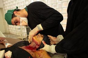
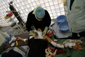
The pain of a severe burn is almost unimaginable -- and so is the destruction it causes. Simply put, your body won't last very long without the skin's protection. Large, open wounds are highly susceptible to bacterial infections and if the body can't regulate its temperature and hydration, it will go into shock. For decades, the best treatment option for a severe burn has been a skin graft.

Advertisement

Skin grafts sound like something straight out of a medieval torture manual, but they save hundreds of thousands of lives every year. More than two million people in the United States require treatment for burns every year and between three and four thousand die from their injuries [source: Merck Manual].

To perform a skin graft, surgeons remove healthy skin from a patient's body and attach it to the wounded area. Extensive scarring is inevitable and the healing process can be long and painful, but the majority of patients will survive the treatment and return to their normal lives.

What are the injuries and conditions that require a skin graft? How is the surgery performed? And what are the latest breakthroughs in artificial skin technology? Keep reading to find out more.

Advertisement

Reasons for a Skin Graft

Doctors prescribe a skin graft when an area of skin is so damaged that it may not be able to regenerate on its own. There are several traumatic injuries, medical conditions and post-surgical procedures that may require skin grafts as part of the treatment process.

Severe burns are some of the most common injuries that are treated with skin grafts. Minor first-degree burns only affect the topmost layer of skin and can usually heal on their own. But if a patient has a second-degree burn that covers more than 10 percent of the body or a third-degree burn that covers more than one percent of the body, it's classified as a severe burn [source: Merck Manual].

Advertisement

What makes these burns so severe? In second-degree burns, the skin damage extends to the dermis, the second layer of skin. If the damaged area is large enough, the burn will have great difficulty closing on its own and will be at high risk of infection. A third-degree burn extends through all three layers of skin, sometimes reaching muscle, cartilage and even bone. Such an extreme injury will almost certainly require a skin graft to heal.

Pressure ulcers, also known as bedsores, are a chronic and dangerous condition that plagues patients who are bedridden or confined to a wheelchair. The ulcers are caused by prolonged pressure on a part of the body, cutting off circulation. Common places for pressure ulcers are the buttocks, hips, back of legs, shoulder blades and even the head. Some pressure ulcers are small, relatively shallow wounds, but a stage IV ulcer -- the most serious kind -- can bore deep enough to damage underlying muscle and bone [source: Mayo Clinic].

Certain types of diabetes can lead to loss of feeling in the feet. This can allow ulcers to form without the patient's knowledge. These ulcers can become deeply infected, eventually leading to amputation. Diabetic ulcers are the most common cause of non-traumatic foot amputation. Skin grafts -- in some cases with lab-generated skin substitutes -- can help cover and close the wound as it heals [source: Scanlan].

Cancer patients often require reconstructive cosmetic surgery to replace skin lost in the removal of cancerous tissue. Breast reconstructions after mastectomies may require that the nipple and areola area be removed and grafted in a new position. If a section of the nose is lost in a skin cancer surgery, doctors can replace it with a skin graft from another part of the body.

Surgeons have developed several different types of skin grafts, depending on the severity of the skin loss. Read more about these techniques in the next section.

Advertisement

Types of Skin Grafts

A doctor performs a skin graft operation on a self-immolation victim in Afghanistan.
Paula Bronstein/Getty Images

In all skin graft procedures, skin must be removed from a donor site and attached to the wounded area. The preferred source of the donor skin is the patient himself. This procedure is called an autograft. Since the body's immune system will ultimately reject foreign skin, autografts are the best option for long-term treatment.

In some cases, the patient might be too sick or injured for doctors to immediately perform an autograft. In that case, doctors may place a temporary skin covering on the affected area to cut down on the chance of infection and buy time until the patient regains his or her strength.

Advertisement

When skin is used from another human (usually a cadaver), it's called an allograft. Allografts can last seven to ten days before the body rejects them [source: WCI]. Xenografts are temporary skin coverings harvested from animals, usually pigs. They only last three to five days [source: BCM]. In some cases, the injury is superficial enough that the patient doesn't need an autograft. The allograft or xenograft can provide enough protection to allow the skin to regenerate on its own.

In cases where an autograft is necessary, there are three major types to choose from: split-thickness grafts, full-thickness grafts and composite grafts. The main criteria for choosing which method to use is the depth of the patient's skin loss. Split-thickness grafts are for the shallowest wounds, or those that affect only the epidermis and part of the dermis. They are called split-thickness grafts because the doctor removes only a few layers of skin from the donor site.

Full-thickness grafts require that all three skin layers -- epidermis, dermis and hypodermis -- be removed from the donor site. Full-thickness grafts are often used in cases where the cosmetic appearance of the injured area is very important, like the face. Full-thickness grafts include hair follicles, sweat glands and blood vessels that allow the graft area to look and function more normally. Split thickness grafts, since they lack underlying glands and blood vessels, tend to look flat and discolored.

A composite graft is for wounds that include bone, tendon, cartilage or the loss of muscle. In the case of a nose reconstruction, for example, the surgeon would need to harvest a composite graft that includes supportive cartilage tissue along with the skin layers.

An exciting new alternative to autografts is lab-generated skin, but we'll talk more about that later. Right now, we're going to walk through the difficult skin graft surgical procedure.

Advertisement

Skin Graft Surgery

For most skin graft procedures, the patient is put under general anesthesia. But if the affected area is very small, the doctor can use a local anesthetic to relieve pain.

The first step is to debride the wound area. In this procedure, the surgeon will meticulously clean the wound area to remove any damaged skin and tissue. He may need to use a scalpel to cut away uneven tissue along the edge of the wound. The goal is to create a clean, disinfected and bleeding surface on which to attach the donor skin.

Advertisement

Next, the surgeon measures the precise area of the wound and traces the identical pattern over the donor site. The donor site can be anywhere on the body, but surgeons usually choose spots that are covered by clothing like the lower back, buttock or inner thigh [source: Medline Plus]

To remove the exact thickness of skin from the donor site, the surgeon uses a special tool called a dermatome. Even though it's a nauseating image, think of a dermatome like a surgical-grade cheese slicer.

To maximize the effectiveness of a minimum amount of donor skin, doctors may choose to mesh the donor skin. To do this, doctors pass the harvested skin layers through a rolling device (resembling an old mimeograph machine) that perforates the skin with hundreds of tiny holes. The result looks like a mesh T-shirt. The surgeon can now stretch the meshed skin to cover a larger wound area. Mesh skin also allows the underlying wound to easily excrete fluids, lessening the risk of infection.

In most cases, the donor skin is placed carefully over the wound and fastened with stitches or surgical staples. For very thin split thickness grafts, it's possible to forgo stitches and simply secure the graft with gauze and dressing. For full-thickness grafts, the graft area will be covered with an antibiotic solution, several layers of mesh gauze as well as extra bandages, elastic netting and even a cast.

The donor site will also require treatment. In the case of a full-thickness graft, the donor site will need to be stitched back together. Split-thickness grafts usually only require lots of antibiotic ointment and clean dressing to regenerate new skin layers.

Skin grafts take time to heal -- this applies to both the donor site and the graft area. And unfortunately, grafts don't always work the first time. Read more about the healing process and possible complications on the next page.

Advertisement

Healing and Complications of Skin Grafts

The healing process for a skin graft operation can last for several weeks, depending on the wound's severity and the source of the skin graft.
Paula Bronstein/Getty Images

Skin cannot survive without oxygen. The best way to infuse skin cells with oxygen and other nutrients is through the blood. Healthy, living skin is full of tiny blood vessels that channel the body's blood supply to grow new skin cells and sustain older ones.

For a skin graft to heal, it must grow and activate new blood vessels. In a successful graft, this regeneration process begins as quickly as 36 hours after surgery [source: Medline Plus].

Advertisement

Because oxygen is so important to the healing process, some doctors prescribe hyperbaric oxygen therapy. Perhaps you've heard of a hyperbaric chamber. It looks like a long, glass-walled tube surrounding a raised bed. Inside a hyperbaric chamber, the patient is exposed to a 100 percent oxygen environment at twice the normal atmospheric pressure. These intense blasts of pure oxygen can speed the healing process of skin grafts.

Another healing technique is something called vacuum-assisted closure (VAC). In this post-operative procedure, the grafted skin is dressed with a porous bandage and attached to a tube that connects to a vacuum source. The vacuum helps draw out interstitial fluids and encourage blood flow to the graft. All potentially infectious fluids are sucked out of the wound for easy disposal. Some surgeons are so impressed with the technique that they leave the VAC tube attached for up to seven days after surgery without even changing the dressing [source: Carson].

The healing process for skin grafts can be slow, depending on the severity of the wound and the size and depth of the donor sites. Patients who receive full-thickness grafts may need to stay in the hospital for as long as two weeks to keep the graft stabilized and infection-free. Split-thickness grafts may only require a few nights at the hospital.

In all cases, patients will have to be very gentle with their graft areas when they get home. They should avoid stretching the skin and abstain from vigorous physical activity for at least a month. Remember that the donor site may take a couple a weeks to heal, too.

All skin grafts leave scars, both in the donor site area and the graft itself. Full-thickness grafts leave a less noticeable scar, because they contain functional blood vessels. Split-thickness grafts, which lack sweat glands, hair follicles and blood vessels, are discolored and need to be moisturized frequently to avoid scaling and chafing.

Unfortunately, some skin grafts can form infections in the area between the donor skin and the wound. Fluid can build up underneath the donor skin, preventing it from successfully attaching to the wound site. In these cases, the graft is said to "fail," and will need to be reattempted with a new batch of donor skin.

All in all, skin grafts can be painful, difficult procedures. The good news is that medical researchers have developed artificial skin products to minimize the use of donor skin and speed the healing process. Read more about lab-grown skin on the next page.

Advertisement

Artificial Skin Grafts

In extreme burn cases, where 80 or 90 percent of a victim's body is scorched by second- and third-degree burns, there often isn't enough healthy donor skin available to perform an autograft. Beginning in the late 1970s, medical researchers began experimenting with sheets of artificial skin that could be permanently grafted onto patients who have no other viable option.

As we explained in the HowStuffWorks.com article, How Lab-grown Skin Works, two Boston surgeons debuted a successful new artificial skin design in 1981 that's now known as Integra. Interestingly, Integra technically isn't "artificial skin." Instead of replicating the function of healthy skin, Integra "tricks" real skin cells to grow in the damaged dermis.

Advertisement

Integra functions as artificial "scaffolding" around which new skin cells can grow. The scaffolding is made of shark cartilage and cow-derived collagen, the protein found in all connective tissue. The bottom layer of the collagen scaffolding -- the part that makes contact with the wound surface -- is covered with a sugar molecule called glycosaminoglycan. The sticky sugar coating mimics the texture of the lower surface of the dermis [source: NIGMS].

Apparently, this texture is enough to trick skin cells in the body called fibroblasts to start generating human collagen on contact. As the body produces more and more collagen, the connective tissue begins to work its way up the artificial scaffolding, slowly building a new dermis. Meanwhile, the artificial scaffolding simply dissolves away, leaving no trace of the implant.

Integra does not replace the epidermis. Instead, the product comes with a thin silicon coating that can be pealed away once the dermis has fully regenerated. To replace the epidermis, doctors rely on lab-grown skin. They examine a piece of skin from the patient and place it in a culture spiked with mouse-derived fibroblasts. Over the course of a few weeks, the fibroblasts generate a thin sheet of epidermis that can be placed over the new dermis.

The latest breakthroughs in artificial skin point to full skin replacements -- epidermis, dermis and even blood vessels -- grown entirely in the lab. A German company has successfully grown bi-layer skin samples (both epidermis and dermis) in a lab and California researchers have successfully grown blood vessels from a skin sample [sources: Maugh and ScienceDaily]. Scientists hope to produce a fully-functional, lab-produced artificial skin graft in the near future.

For lots more information on amazing medical procedures and the science of skin, take a look at the links on the next page.

Advertisement

Lot More Information

Related HowStuffWorks Articles

More Great Links

Sources

  • Baylor College of Medicine. "Skin Graft" https://www.thehearthospitalbaylor.com/handler.cfm?event=practice,template&cpid=53325
  • Calman, Barney. "The burns victim saved by £60K of 'shark skin.'" The Daily Mail. September 14, 2009 http://www.dailymail.co.uk/health/article-1213003/The-burns-victim-saved-60K-shark-skin.html
  • Carson, Stanley N. et al. "Vacuum-Assisted Closure Used for Healing Chronic Wounds and Skin Grafts in the Lower Extremities." Ostomy Wound Management. Volume 50: Issue 3 (October 13, 2009) http://www.o-wm.com/article/2386
  • The Massachusetts General Hospital Burn Research Center. "The Development of Artificial Skin" http://www.burnresearchcenter.org/brcpublicwebsite/artificialskin.htm
  • Maugh, Thomas H. "Kidney patient's cells used to grow blood vessels in lab." Los Angeles Times. April 25, 2009 http://articles.latimes.com/2009/apr/25/science/sci-kidney25
  • Mayo Clinic. "Bedsores (pressure sores)" http://www.mayoclinic.com/health/bedsores/DS00570
  • Medline Plus. "Skin graft" http://www.nlm.nih.gov/medlineplus/ency/article/002982.htm
  • The Merck Manual Online Medical Library. "Burns" http://www.merck.com/mmhe/sec24/ch289/ch289a.html
  • National Institute of General Medical Sciences. "Artificial Skin Fact Sheet" http://www.ncbi.nlm.nih.gov/pmc/articles/PMC3021617/
  • Norfolk and Norwich University Hospitals. "Split Thickness Skin Graft." http://www.nnuh.nhs.uk/docs%5Cdocuments%5C37.htm
  • Scanlan, Matt. "New skin graft eases diabetic foot ulcers." November 21, 1997 http://www.mc.vanderbilt.edu/reporter/index.html?ID=350
  • Science Daily. "Artifical Skin Manufactured in Fully Automated Process." May 19, 2009 http://www.sciencedaily.com/releases/2009/05/090518102959.htm
  • Swedish Medical Center. "Skin Graft" http://healthlibrary.epnet.com/GetContent.aspx?token=af362d97-4f80-4453-a175-02cc6220a387&chunkiid=14862
  • Indian Journal of Plastic Surgery. "Biologic and Synthetic Skin Substitutes: An overview" http://www.ncbi.nlm.nih.gov/pmc/articles/PMC3038402/

Advertisement

Advertisement

Loading...