How Nerves Work

Nerves (green) and arteries (red) in the embryonic limb skin. See more brain pictures.
Image courtesy National Institutes of Health

Consider this. You touch a hot object and immediately drop it or withdraw your hand from the heat source. You do this so quickly you don't even think about it. How does this happen? Your nervous system coordinated everything. It sensed the hot object and signaled your muscles to let it go. Your nervous system, which consists of your brain, spinal cord, peripheral nerves and autonomic nerves, coordinates all movements, thoughts and sensations that you have. In this article, we'll examine the structure and functions of your nervous system, how nerve cells communicate with each other and various tissues and what can go wrong when nerves become damaged or diseased.

The nervous system:

Advertisement

  • Senses your external and internal surroundings
  • Communicates information between your brain and spinal cord and other tissues
  • Coordinates voluntary movements
  • Coordinates and regulates involuntary functions like breathing, heart rate, blood pressure and body temperature.

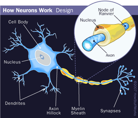
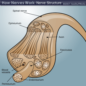
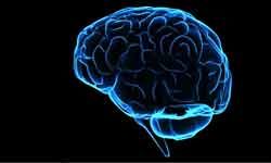
The brain is the center of the nervous system, like the microprocessor in a computer. The spinal cord and nerves are the connections, like the gates and wires in the computer. Nerves carry electrochemical signals to and from different areas of the nervous system as well as between the nervous system and other tissues and organs. Nerves are divided into four classes:

  1. Cranial nerves connect your sense organs (eyes, ears, nose, mouth) to your brain
  2. Central nerves connect areas within the brain and spinal cord
  3. Peripheral nerves connect the spinal cord with your limbs
  4. Autonomic nerves connect the brain and spinal cord with your organs (heart, stomach, intestines, blood vessels, etc.)

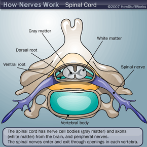
The central nervous system consists of the brain and spinal cord, including cranial and central nerves. The peripheral nervous system consists of the peripheral nerves, and the autonomic nervous system is made of autonomic nerves. Fast reflexes, like removing your hand quickly from a heat source, involve peripheral nerves and the spinal cord. Thought processes and autonomic regulation of your organs involve various parts of the brain and are relayed to the muscles and organs through the spinal cord and peripheral/autonomic nerves.

Advertisement

The Spinal Cord and Neurons

The spinal cord extends through hollow openings in each vertebra in your back. It contains various nerve cell bodies (gray matter) and nerve processes or axons (white matter) that run to and from the brain and outward to the body. The peripheral nerves enter and exit through openings in each vertebra. Within the vertebra, each nerve separates into dorsal roots (sensory nerve cell processes and cell bodies) and ventral roots (motor nerve cell processes). The autonomic nerve cell bodies lie along a chain that runs parallel with the spinal cord and inside the vertebrae, while their axons exit in the spinal nerve sheaths.

Nerve Cells

The brain, spinal cord and nerves consist of more than 100 billion nerve cells, called neurons. Neurons gather and transmit electrochemical signals. They have the same characteristics and parts as other cells, but the electrochemical aspect lets them transmit signals over long distances (up to several feet or a few meters) and pass messages to each other.

Advertisement

2007 HowStuffWorks

Neurons have three basic parts:

  • Cell body: This main part has all of the necessary components of the cell, such as the nucleus (which contains DNA), endoplasmic reticulum and ribosomes (for building proteins) and mitochondria (for making energy). If the cell body dies, the neuron dies. Cell bodies are grouped together in clusters called ganglia, which are located in various parts of the brain and spinal cord.
  • Axons: These long, thin, cable-like projections of the cell carry electrochemical messages (nerve impulses or action potentials) along the length of the cell. Depending upon the type of neuron, axons can be covered with a thin layer of myelin, like an insulated electrical wire. Myelin is made of fat, and it helps to speed transmission of a nerve impulse down a long axon. Myelinated neurons are typically found in the peripheral nerves (sensory and motor neurons), while nonmyelinated neurons are found within the brain and spinal cord.
  • Dendrites or nerve endings: These small, branchlike projections of the cell make connections to other cells and allow the neuron to talk with other cells or perceive the environment. Dendrites can be located on one or both ends of the cell.

Neurons come in many sizes. For example, a single sensory neuron from your fingertip has an axon that extends the length of your arm, while neurons within the brain may extend only a few millimeters. Neurons have different shapes depending on what they do. Motor neurons that control muscle contractions have a cell body on one end, a long axon in the middle and dendrites on the other end; sensory neurons have dendrites on both ends, connected by a long axon with a cell body in the middle.

Neurons also vary with respect to their functions:

  • Sensory neurons carry signals from the outer parts of your body (periphery) into the central nervous system.
  • Motor neurons (motoneurons) carry signals from the central nervous system to the outer parts (muscles, skin, glands) of your body.
  • Receptors sense the environment (chemicals, light, sound, touch) and encode this information into electrochemical messages that are transmitted by sensory neurons.
  • Interneurons connect various neurons within the brain and spinal cord.

In peripheral and autonomic nerves, the axons get bundled into groups, based on where they're coming from and going to. The bundles are covered by various membranes (fasciculi). Tiny blood vessels travel through the nerves to supply the tissues with oxygen and remove waste. Most peripheral nerves travel near major arteries deep within limbs and close to the bones.

Next, we'll learn about neural pathways.

Advertisement

Neural Pathways and Action Potentials

Neural pathways

The simplest type of neural pathway is a monosynaptic (single connection) reflex pathway, like in the knee-jerk reflex. When the doctor taps a certain spot on your knee with a rubber hammer, receptors send a signal into the spinal cord through a sensory neuron. The sensory neuron passes the message to a motor neuron that controls your leg muscles. Nerve impulses travel down the motor neuron and stimulate the appropriate leg muscle to contract. Nerve impulses also travel to the opposing leg muscle to inhibit contraction so that it relaxes (this pathway involves interneurons). The response is a quick muscular jerk that does not involve your brain. Humans have lots of hardwired reflexes like this, but as tasks become more complex, the pathway "circuitry" gets more complicated and the brain gets involved.

­­Action potentials

Advertisement

We have talked about nerve signals and mentioned that they are electrochemical in nature, but what does that mean?

To understand how neurons transmit signals, we must first look at the structure of the cell membrane. The cell membrane is made of fats or lipids called phospholipids. Each phospholipid has an electrically charged head that sticks near water and two polar tails that avoid water. The phospholipids arrange themselves in a two-layer lipid sandwich with the polar heads sticking into water and the polar tails sticking near each other. In this configuration, they form a barrier that separates the inside of the cell from the outside and that does not permit water-soluble or charged particles (like ions) from moving through it.

So how do charged particles get into cells? We'll find out on the next page.

Advertisement

Ion Channels

© Photographer: Eraxion | Agency: Dreamstime

Because ions are charged and water-soluble, they must move through small tunnels or channels (specialized proteins) that span the cell membrane's lipid bilayer. Each channel is specific for only one type of ion. There are specific channels for sodium ions, potassium ions, calcium ions and chloride ions. These channels make the cell membrane selectively permeable to various ions and other substances (like glucose). The selective permeability of the cell membrane allows the inside to have a different composition than the outside.

For the purposes of nerve signals, we are interested in the following characteristics:

Advertisement

  • The outside fluid is rich in sodium, a concentration about 10 times higher than the inside fluid
  • The inside fluid is rich in potassium, a concentration about 20 times higher inside the cell than outside.
  • There are large negatively charged proteins inside the cell that are too big to move across the membrane. They give the inside of the cell a negative electrical charge compared to the outside. The charge is about 70 to 80 millivolts (mV) -- 1 mV is 1/1000th of a volt. For comparison, the charge in your house is about 120 V, about 1.2 million times more.
  • The cell membrane is slightly "leaky" to sodium and potassium ions, so a sodium-potassium pump is located in the membrane. This pump uses energy (ATP) to pump sodium ions from the inside to the outside and potassium ions from the outside to the inside.
  • Because sodium and potassium ions are positively charged, they carry tiny electrical currents when they move across the membrane. If sufficient numbers move across the membrane, you can measure the electrical currents.

Nerve Signals

The nerve signal, or action potential, is a coordinated movement of sodium and potassium ions across the nerve cell membrane. Here's how it works:

  1. As we discussed, the inside of the cell is slightly negatively charged (resting membrane potential of -70 to -80 mV).
  2. A disturbance (mechanical, electrical, or sometimes chemical) causes a few sodium channels in a small portion of the membrane to open.
  3. Sodium ions enter the cell through the open sodium channels. The positive charge that they carry makes the inside of the cell slightly less negative (depolarizes the cell).
  4. When the depolarization reaches a certain threshold value, many more sodium channels in that area open. More sodium flows in and triggers an action potential. The inflow of sodium ions reverses the membrane potential in that area (making it positive inside and negative outside -- the electrical potential goes to about +40 mV inside)
  5. When the electrical potential reaches +40 mV inside (about 1 millisecond later), the sodium channels shut down and let no more sodium ions inside (sodium inactivation).
  6. The developing positive membrane potential causes potassium channels to open.
  7. Potassium ions leave the cell through the open potassium channels. The outward movement of positive potassium ions makes the inside of the membrane more negative and returns the membrane toward the resting membrane potential (repolarizes the cell).
  8. When the membrane potential returns to the resting value, the potassium channels shut down and potassium ions can no longer leave the cell.
  9. The membrane potential slightly overshoots the resting potential, which is corrected by the sodium-potassium pump, which restores the normal ion balance across the membrane and returns the membrane potential to its resting level.
  10. Now, this sequence of events occurs in a local area of the membrane. But these changes get passed on to the next area of membrane, then to the next area, and so on down the entire length of the axon. Thus, the action potential (nerve impulse or nerve signal) gets transmitted (propagated) down the nerve cell.­

There are a few things to note about the propagation of the action potential.

Advertisement

­When an area has been depolarized and repolarized and the action potential has moved on to the next area, there is a short period of time before that first area can be depolarized again (refractory period). This refractory period prevents the action potential from moving backward and keeps everything moving in one direction.

  • The action potential is an "all-or-none" response. Once the membrane reaches a threshold, it will depolarize to +40 mV. In other words, once the ionic events are set in motion, they will continue until the end.
  • These ionic events occur in many excitable cells besides neurons (like muscle cells).
  • Action potentials are propagated rapidly. Typical neurons conduct at 10 to 100 meters per second. Conduction speed varies with the diameter of the axon (larger = faster) and the presence of myelin (myelinated = faster). The rapid nerve conductions throughout the neural circuitry enable you to respond to stimuli in fractions of a second.
  • The channels can be poisoned and prevented from opening. Various toxins (puffer fish toxin, snake venom, scorpion venom) can prevent specific channels from opening and distort the action potential or prevent it from happening altogether. Similarly, many local anesthetics (e.g. lidocaine, novocaine, benzocaine) can prevent action potentials from being propagated in the nerve cells in an area and temporarily prevent you from feeling pain.
  • The propagation of the action potential is also sensitive to temperature in experimental settings. Colder temperatures slow down the action potential, but this usually doesn't happen in an individual. However, you can use cold-block techniques to temporarily anesthetize an area (like putting ice on an injured finger).

So, if the size of the action potential does not vary, how does an action potential code information? Information is encoded by the frequency of action potentials, much like FM radio. A small stimulus will initiate a low frequency train of a few action potentials. As the intensity of the stimulus increases, so does the frequency of action potentials.

On the next page, we'll learn about how nerves communicate with each other.

Advertisement

Synaptic Transmission

Like wires in your home's electrical system, nerve cells make connections with one another in circuits called neural pathways. Unlike wires in your home, nerve cells do not touch, but come close together at synapses. At the synapse, the two nerve cells are separated by a tiny gap, or synaptic cleft. The sending neuron is called the presynaptic cell, while the receiving one is called the postsynaptic cell. Nerve cells send chemical messages with neurotransmitters in a one-way direction across the synapse from presynaptic cell to postsynaptic cell.

Let's look at this process in a neuron that uses the neurotransmitter serotonin:

Advertisement

  1. The presynaptic cell (sending cell) makes serotonin (5-hydroxytryptamine, 5HT) from the amino acid tryptophan and packages it in vesicles in its end terminals.
  2. An action potential passes down the presynaptic cell into its end terminals.
  3. Serotonin passes across the synaptic cleft, binds with special proteins called receptors on the membrane of the postsynaptic cell (receiving cell) and sets up a depolarization in the postsynaptic cell. If the depolarizations reach a threshold level, a new action potential will be propagated in that cell. Some neurotransmitters cause the postsynaptic cell to hyperpolarize (the membrane potential becomes more negative, which would inhibit the formation of action potentials in the postsynaptic cell). Serotonin fits with its receptor like a lock and key.
  4. The remaining serotonin molecules in the cleft and those released by the receptors after use get destroyed by enzymes in the cleft (monoamine oxidase (MAO), catechol-o-methyl transferase (COMT)). Some get taken up by specific transporters on the presynaptic cell (reuptake). In the presynaptic cell, MAO and COMT destroy the absorbed serotonin molecules. This enables the nerve signal to be turned "off" and readies the synapse to receive another action potential.
  5. There are several types of neurotransmitters besides serotonin, including acetylcholine, norepinephrine, dopamine and gamma-amino butyric acid (GABA). Any given neuron produces only one type of neurotransmitter. Any one nerve cell may have synapses on it from excitatory presynaptic neurons and from inhibitory presynaptic neurons. In this way, the nervous system can turn various cells (and subsequent neural pathways) "on" and "off." Finally, nerve cells synapse on effector cells (muscles, glands, etc.) to evoke or inhibit responses.

Next, we'll learn about the different types of sensory neurons.

­

Advertisement

Sensory Neurons

The nervous system has many types of sensory neurons. Nerve endings on one end of each neuron are encased in a special structure to sense a specific stimulus.

  • Chemoreceptors sense chemicals. The olfactory bulb that monitors your sense of smell has chemoreceptors that sense odors (chemicals in the air). Taste buds have chemoreceptors to detect chemicals dissolved in liquids. Chemoreceptors in the brain also monitor the concentration of carbon dioxide in the blood and cerebrospinal fluid to help control your rate of breathing.
  • Mechanoreceptors sense touch, pressure and distortion (stretch). Stretch receptors in your muscle tendons are the first link in the knee-jerk reflex.
  • Photoreceptors, which sense light, are found in the retinas of your eyes.
  • Thermoreceptors are free nerve endings that sense temperature, but we're not sure exactly how they do this. Changes in temperature could affect the movements of ions across the cell membrane and influence action potentials in that way.
  • Nociceptors are free nerve endings that sense pain. They respond to a variety of stimuli (heat, pressure, chemicals) and sense tissue damage.
  • Auditory receptors in the inner ear sense vibrations from sound waves.

Typically, a stimulus causes ionic changes in the receptor neuron's dendrites, which lead to the formation of action potentials in the receptor neurons. These action potentials travel the sensory neuron, which connects to a motor neuron (and possibly an ascending neuron) in the spinal cord. The action potential causes neurotransmitter release within the presynaptic cell. The neurotransmitter binds to the postsynaptic cell and elicits an action potential there. The action potential will travel the length of the postsynaptic cell to another synapse on the effector cell (like a muscle cell, skin, blood vessel, gland), where its neurotransmitter will cause a response in the effector cell (like a muscle contraction). Alternatively, the postsynaptic cell may be another neuron that transmits the signal to another neuron in the brain or spinal cord.

Advertisement

What happens when nerves are damaged or diseased? We'll find out on the next page.

Advertisement

Nerve Disorders

Nerve activity can be affected by toxins, trauma and disease.

  • Toxic substances interfere with sodium or potassium channels, whose actions underlie the action potential. Such toxic substances include venoms, heavy metals (like mercury and lead) and anesthetics.
  • Trauma happens when limbs or vertebrae become fractured and the nerves close to them are crushed, pinched or even severed. This can result in pain, numbness, complete loss of feeling or loss of movement. The extent of damage and recovery depends upon the severity and location of the injury.
  • A pinched nerve is a common problem in which a bone, joint or muscle compresses a nerve and impairs its conduction, leading to pain and numbness. This often occurs between vertebrae in the spine, where swelling discs can compress the nerves as they exit.
  • Another common example is carpal tunnel syndrome, where repeated motions of the wrist (like from typing on computers) causes swelling in the bone tunnel (carpal bones) where the radial and ulnar nerves pass through the wrist into the fingers. Sciatica is a similar nerve problem where an injured spinal disc compresses the sciatic nerve to the leg, causing pain and numbness.
  • Some diseases directly affect nerve function. For example, multiple sclerosis (MS) occurs when the myelin surrounding nerves degrades, which affects nerve conduction. MS may be caused by an autoimmune response, where the patient's own immune system attacks the myelinated nerves. Myasthenia gravis (MG) is a disease in which the synaptic transmission between nerve cells and muscle cells is disrupted.

Your nerves must conduct impulses correctly for you to regulate your internal environment, respond to your external environment, think, and learn. When nerves become impaired, many body functions or quality of life can be affected.

Advertisement

To learn more about nerves, check out the links on the following page.

Advertisement

Lots More Information

Related Articles

More Great Links

  • Excitable Cells. http://users.rcn.com/jkimball.ma.ultranet/BiologyPages/E/ExcitableCells.html
  • Getting Up the Nerve, Chicago Tribune online lesson. http://nie.chicagotribune.com/activities_082205.htm
  • HHMI BioInteractive Neuroscience Virtual Lab. http://www.hhmi.org/biointeractive/neuroscience/vlab.html
  • Neuroscience for Kids. http://faculty.washington.edu/chudler/introb.html
  • New York Times, "The Other Brain Also Deals With Many Woes." http://www.nytimes.com/2005/08/23/health/23gut.html
  • NIGMS/NIG. "Inside the Cell." http://publications.nigms.nih.gov/insidethecell/index.html
  • NIH, NIDA curriculum The Brain: Understanding Neurobiology Through the Study of Addiction. http://science-education.nih.gov/supplements/nih2/addiction/default.htm
  • NLM/NIH Medline Plus, Multiple Sclerosis. http://www.nlm.nih.gov/medlineplus/multiplesclerosis.html
  • NLM/NIH Medline Plus, Myasthenia Gravis. http://www.nlm.nih.gov/medlineplus/myastheniagravis.html.
  • NLM/NIH Medline Plus, Nerve Conduction Velocity. http://www.nlm.nih.gov/medlineplus/ency/article/003927.htm.
  • Online Etymology Dictionary: Nerve. http://www.etymonline.com/index.php?l=n&p=3
  • Psychology Today, "Our Second Brain: the Stomach." http://psychologytoday.com/articles/pto-19990501-000013.html
  • Purves, D et al, "Neuroscience Chapter 1: Nerve cells." http://www.ncbi.nlm.nih.gov/books/bv.fcgi?indexed=google&rid=neurosci.section.50
  • Society for Neuroscience, Astrocytes. http://www.sfn.org/index.cfm?pagename=brainBriefings_adult_neurogenesis
  • Society for Neuroscience, Axon Guidance. http://www.sfn.org/index.cfm?pagename=brainBriefings_axonGuidance
  • Society for Neuroscience, Blood-Brain Barrier. http://www.sfn.org/index.cfm?pagename=brainBriefings_bloodBrainBarrier
  • Society for Neuroscience, Brain Backgrounders, How Do Nerve Cells Communicate? http://www.sfn.org/index.cfm?pagename=brainBackgrounders_howDoNerveCells Communicate
  • Society for Neuroscience, Brain Briefings: Adult Neurogenesis. http://www.sfn.org/index.cfm?pagename=brainBriefings_adult_neurogenesis
  • Society for Neuroscience, Myelin and Spinal Cord Repair. http://www.sfn.org/index.cfm?pagename=brainBriefings_myelinAndSpinalCordRepair
  • Society for Neuroscience, Neuron Migration and Brain Disorders. http://www.sfn.org/index.cfm?pagename=brainBriefings_neuron MigrationAndBrainDisorders
  • Society for Neuroscience, Neurotrophic Factors. http://www.sfn.org/index.cfm?pagename=brainBriefings_neurotrophicFactors
  • Society for Neuroscience, Nociceptors and Pain. http://www.sfn.org/index.cfm?pagename=brainBriefings_nociceptorsAndPain
  • Society for Neuroscience, Spinal Cord Repair. http://www.sfn.org/index.cfm?pagename=brainBriefings_spinalCordRepair
  • Society for Neuroscience. Brain Facts. http://www.sfn.org/index.cfm?pagename=brainFacts

Advertisement

Loading...