5 Inherited Skin Problems

psoriasis
Some genodermatoses, like psoriasis, can be severe, but in no way life threatening. Others can be fatal. See more pictures of skin problems.
iStockphoto/Thinkstock

Despite its comparative toughness and impressive size, many problems can plague the skin. Ultraviolet radiation from the sun can damage the DNA in skin cells, leading to sunburn or even cancer. The effects of dry and windy climates can wick moisture from skin, leading to chapped, painful areas. Sebaceous glands can become clogged, which can cause pimples.

There are certainly measures we can take to prevent damage to our skin from environmental exposure. But there are also skin disorders that take place from the inside and are the result of genetic mutations that we can inherit from our parents. These inherited skin problems are collectively referred to as genodermatoses, or genetic dermatological disorders.

Advertisement

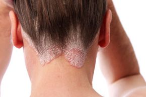
The range of genodermatoses is sweeping. Some, like psoriasis, can be relatively mild and treatable with topical remedies. Others, like harlequin-type ichthyosis, can be fatal. What follows are five inherited skin problems that cover a lot of epidermis.

5: Darier-White Disease

fingernails
The same process that builds fingernails can also take place elsewhere on the skin in a condition known as Darier-White disease.
Pixland/Thinkstock

Darier-White disease, known clinically as keratosis follicularis, was first identified in the late 19th century by dermatologists Ferdinand-Jean Darier and James Clarke White. White identified the disorder as an inherited skin condition when a mother and daughter came to him for treatment.

Darier-White disease is characterized by abnormal hardening of skin cells on the outer layer of the skin, a process called keratinization. This is the same process that converts healthy skin cells into fingernails, but Darier-White disease sufferers have this occur elsewhere on the skin. Researchers have found that the skin cells of sufferers lack the junction that binds cells together (called desmosomes) which may allow keratinization to take place more frequently [source: Kwok]. The most visible symptom is small, greasy, yellow-brown, pimplelike papules that form en masse in areas around sebaceous glands [source: Spitz].

Advertisement

Researchers have found links between ATP2A2 gene mutations and the Darier-White disease, but have also found no common mutation pattern that defines the condition. Even people with the same ATP2A2 mutations can present different symptoms.

4: Epidermolysis Bullosa

blister
bullosa leads to painful blisters from slight temperature and pressure.
©iStockphoto/whitemay

Epidermolysis bullosa (EB) is a debilitating inherited skin condition that leads to blistering from mild pressure or temperature changes. Normally humans have a blister response when the outer and inner layers of skin become separated. The loose area becomes filled with fluid that serves as a cushion while the injured skin beneath heals.

Patients with epidermolysis bullosa have an extremely heightened blister response. Simply walking, crawling, being held and or experiencing even slight changes in room temperature can lead to painful blistering across the skin. The frequency of blistering increases the chance the patient will suffer an infection, leading to further health risks like amputation.

Advertisement

At least 10 different genes have been linked in combination to produce EB. In most cases, the condition is inherited from the parents, though it can also rarely result from a random mutation [source: Mayo Clinic]. Most commonly, a mutation on genes responsible for expressing keratin proteins that provide structure and strength for the binding between skin layers is inherited from the parents [source: Marinkovich].

3: Lamellar Ichthyosis

skin cell
In healthy skin, newer skin cells replace old ones, which shed. Patients with lamellar ichthyosis produce healthy skin cells but lack the ability to shed them, leading to scaly plates across their bodies.
Hemera/Thinkstock

The inherited condition lamellar ichthyosis gets the second half of its name from the Latin word for fish, ichthys. The term is appropriate since patients with lamellar ichthyosis develop thick scales all over as a result of their disorder.

In healthy people, old cells protect young cells before shedding and being replaced. Beneath the stratum corneum (outer layer), skin cells known as keratinocytes divide to form healthy new cells. As they age and die, keratinocytes harden and migrate toward the stratum corneum to form a protective barrier. Eventually, they're shed as newly hardened cells replace them. In lamellar ichthyosis, the skin cells of the patient develop normally, but as they harden and migrate to the stratum corneum they don't separate, which prevents them from shedding. Eventually the dead skin cells build up and form the hard, scaly plates that cover the body and characterize lamellar ichthyosis [source: WebMD].

Advertisement

The condition is rare. Generally, patients develop symptoms in early childhood or may be born with scales. The symptoms may disappear during most of adulthood and reappear later in life [source: Mayo Clinic].

2: Cutaneous Porphyria

red blood cells
The inability to convert porphyrins into heme lies beneath the inherited skin condition of cutaneous porphyria.
iStockphoto/Thinkstock

Cutaneous porphyrias actually make up six different types of the inherited disorder porphyria. In each, patients cannot produce enzymes that create heme, a component of red blood cells that delivers oxygen. Heme is made of chemicals called porphyrins, and they can accumulate when they aren't converted into heme. This lack of heme and accumulation of porphyrins lead to the symptoms of porphyria. Cutaneous porphyrias target the skin.

In cases of cutaneous porphyria, the patient's skin is extremely photosensitive. The skin develops redness, painful irritation and blisters after being exposed to sunlight for a very short period of time. The skin may also swell when exposed to sunlight, and can also darken abnormally [source: American Porphyria Foundation]. As a result, patients are strongly advised to avoid exposure to sunlight.

Advertisement

Children inherit the disorder from one or more of their parents. There are eight different enzymes that convert porphyrins to heme, and an inherited mutation on any of the genes that express these proteins can lead to porphyria.

1: Mal de Maleda

croatia
Mal de Maleda was named after the island of Maleda, off the coast of Croatia, where the condition was first documented.
Adam Jones/Getty Images

As far as genodermatoses go, Mal de Maleda is one of the rarer types. This inherited skin disorder is found mainly among people of Mediterranean descent, and was named for the island of Maleda, located near Croatia, where the first cases were documented [source: Goldsmith].

The disorder is a variation of keratosis palmaris et plantaris, a more prevalent type of skin disorder characterized by a thickening of the skin on the palms of the hands and the soles of the feet. This increase in size is the result of an enlargement in the size of the skin cells. This, in turn, leads to an enlargement of the palms and the soles, and also gives them a yellowish hue [source: Wrong Diagnosis].

Advertisement

Since Mal de Maleda is a rare genodermatosis, it's not surprising that it's also an autosomal recessive disorder. This means that two copies of the mutated gene must be contributed, one from each parent.

The SLURP1 gene, which is responsible for encoding the proteins that form the binding between cells, has been identified as the culprit behind Mal de Maleda [source: Spitz].

Lots More Information

Related Articles

  • American Porphyria Foundation. "Porphyria cutanea tarda (PCT)." Accessed May 25, 2010.http://www.porphyriafoundation.com/about-porphyria/types-of-porphyria/PCT
  • Goldsmith, Lowell A. "Slurping across the mare nostrum." Journal of Investigative Dermatology. 2003.http://www.nature.com/jid/journal/v120/n3/full/5603368a.html
  • Kwok, Pui-Yan, MD, PhD. "Keratosis follicularis (Darier disease)." eMedicine. May 14, 2010.http://emedicine.medscape.com/article/1107340-overview
  • Macnair, Dr. Trisha. "What is porphyria?" Accessed May 25, 2010.http://www.bbc.co.uk/health/physical_health/conditions/porphyria1.shtml
  • Marinkovich, M Peter. "Epidermolysis bullosa." eMedicine. November 30, 2009.http://emedicine.medscape.com/article/1062939-overview
  • Mayo Clinic. "Epidermolysis bullosa." September 26, 2009http://www.mayoclinic.com/health/epidermolysis-bullosa/DS01015.
  • Mayo Clinic. "Ichthyosis vulgaris." April 8, 2010.http://www.mayoclinic.com/health/ichthyosis-vulgaris/DS00734/DSECTION%3Dsymptoms
  • Mayo Clinic. "Porphyria." April 8, 2010.http://www.mayoclinic.com/health/porphyria/DS00955
  • National Cancer Institute. "Study of scaling disorders and other inherited skin diseases." Clinical
  • Trials.gov. November 30, 1999.http://clinicaltrials.gov/ct2/show/NCT00001292
  • Spitz, Joel H. "Genodermatoses: a clinical guide to genetic skin disorders."
  • The Biology Project. "Cell membranes problem set." University of Arizona. August 2004.http://www.biology.arizona.edu/cell_bio/problem_sets/membranes/13t.html
  • WebMD. "Ichthyosis, lamellar." March 9, 2010.http://www.webmd.com/skin-problems-and-treatments/ichthyosis-lamellar-nord
  • Wrong Diagnosis. "Keratosis palmaris et plantaris." Accessed May 25, 2010.http://www.wrongdiagnosis.com/medical/keratosis_palmaris_et_plantaris.htm

Advertisement

Loading...